8AJN

Structure of the human DDB1-DCAF12 complex


Experimental Data Snapshot

  • Method: ELECTRON MICROSCOPY
  • Resolution: 3.00 Å
  • Aggregation State: PARTICLE 
  • Reconstruction Method: SINGLE PARTICLE 

wwPDB Validation   3D Report Full Report


This is version 1.2 of the entry. See complete history


Literature

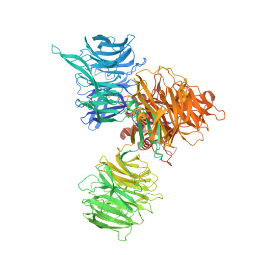
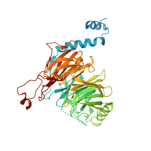
Recognition of the CCT5 di-Glu degron by CRL4 DCAF12 is dependent on TRiC assembly.

Pla-Prats, C.Cavadini, S.Kempf, G.Thoma, N.H.

(2023) EMBO J 42: e112253-e112253

  • DOI: https://doi.org/10.15252/embj.2022112253
  • Primary Citation of Related Structures:  
    8AJM, 8AJN, 8AJO

  • PubMed Abstract: 

    Assembly Quality Control (AQC) E3 ubiquitin ligases target incomplete or incorrectly assembled protein complexes for degradation. The CUL4-RBX1-DDB1-DCAF12 (CRL4 DCAF12 ) E3 ligase preferentially ubiquitinates proteins that carry a C-terminal double glutamate (di-Glu) motif. Reported CRL4 DCAF12 di-Glu-containing substrates include CCT5, a subunit of the TRiC chaperonin. How DCAF12 engages its substrates and the functional relationship between CRL4 DCAF12 and CCT5/TRiC is currently unknown. Here, we present the cryo-EM structure of the DDB1-DCAF12-CCT5 complex at 2.8 Å resolution. DCAF12 serves as a canonical WD40 DCAF substrate receptor and uses a positively charged pocket at the center of the β-propeller to bind the C-terminus of CCT5. DCAF12 specifically reads out the CCT5 di-Glu side chains, and contacts other visible degron amino acids through Van der Waals interactions. The CCT5 C-terminus is inaccessible in an assembled TRiC complex, and functional assays demonstrate that DCAF12 binds and ubiquitinates monomeric CCT5, but not CCT5 assembled into TRiC. Our biochemical and structural results suggest a previously unknown role for the CRL4 DCAF12 E3 ligase in overseeing the assembly of a key cellular complex.


  • Organizational Affiliation

    Friedrich Miescher Institute for Biomedical Research, Basel, Switzerland.


Macromolecules
Find similar proteins by:  (by identity cutoff)  |  3D Structure
Entity ID: 1
MoleculeChains Sequence LengthOrganismDetailsImage
DNA damage-binding protein 11,164Homo sapiensMutation(s): 0 
Gene Names: DDB1XAP1
UniProt & NIH Common Fund Data Resources
Find proteins for Q16531 (Homo sapiens)
Explore Q16531 
Go to UniProtKB:  Q16531
PHAROS:  Q16531
GTEx:  ENSG00000167986 
Entity Groups  
Sequence Clusters30% Identity50% Identity70% Identity90% Identity95% Identity100% Identity
UniProt GroupQ16531
Sequence Annotations
Expand
  • Reference Sequence
Find similar proteins by:  (by identity cutoff)  |  3D Structure
Entity ID: 2
MoleculeChains Sequence LengthOrganismDetailsImage
DDB1- and CUL4-associated factor 12477Homo sapiensMutation(s): 0 
Gene Names: DCAF12KIAA1892TCC52WDR40A
UniProt & NIH Common Fund Data Resources
Find proteins for Q5T6F0 (Homo sapiens)
Explore Q5T6F0 
Go to UniProtKB:  Q5T6F0
PHAROS:  Q5T6F0
GTEx:  ENSG00000198876 
Entity Groups  
Sequence Clusters30% Identity50% Identity70% Identity90% Identity95% Identity100% Identity
UniProt GroupQ5T6F0
Sequence Annotations
Expand
  • Reference Sequence
Experimental Data & Validation

Experimental Data

  • Method: ELECTRON MICROSCOPY
  • Resolution: 3.00 Å
  • Aggregation State: PARTICLE 
  • Reconstruction Method: SINGLE PARTICLE 
EM Software:
TaskSoftware PackageVersion
RECONSTRUCTIONRELION
MODEL REFINEMENTRosetta
MODEL REFINEMENTPHENIX

Structure Validation

View Full Validation Report


Entry History & Funding Information

Deposition Data


Funding OrganizationLocationGrant Number
European Research Council (ERC)European Union--

Revision History  (Full details and data files)

  • Version 1.0: 2022-11-09
    Type: Initial release
  • Version 1.1: 2022-12-21
    Changes: Database references
  • Version 1.2: 2023-05-24
    Changes: Database references