


Structure of iRFP variant C15S/N136R/V256C in complex with phycocyanobilin
Remeeva, A., Kovalev, K., Gushchin, I., Fonin, A., Turoverov, K., Stepanenko, O.To be published.
Experimental Data Snapshot
Starting Model: experimental
View more details
Entity ID: 1 | |||||
|---|---|---|---|---|---|
| Molecule | Chains | Sequence Length | Organism | Details | Image |
| Near-infrared fluorescent protein | 316 | Rhodopseudomonas palustris CGA009 | Mutation(s): 3 Gene Names: iRFP | 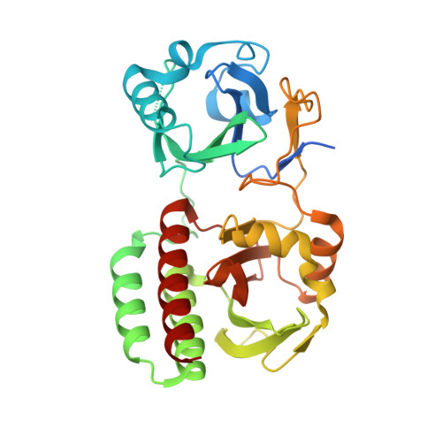 | |
UniProt | |||||
Entity Groups | |||||
| Sequence Clusters | 30% Identity50% Identity70% Identity90% Identity95% Identity100% Identity | ||||
| UniProt Group | G1FNL7 | ||||
Sequence AnnotationsExpand | |||||
Reference Sequence | |||||
| Ligands 1 Unique | |||||
|---|---|---|---|---|---|
| ID | Chains | Name / Formula / InChI Key | 2D Diagram | 3D Interactions | |
| CYC (Subject of Investigation/LOI) Download:Ideal Coordinates CCD File | C [auth A], D [auth B] | PHYCOCYANOBILIN C33 H40 N4 O6 VXTXPYZGDQPMHK-GMXXPEQVSA-N |  | ||
| Length ( Å ) | Angle ( ˚ ) |
|---|---|
| a = 77.806 | α = 90 |
| b = 98.708 | β = 90 |
| c = 156.833 | γ = 90 |
| Software Name | Purpose |
|---|---|
| BUSTER | refinement |
| PDB_EXTRACT | data extraction |
| XDS | data reduction |
| STARANISO | data scaling |
| MOLREP | phasing |
| Funding Organization | Location | Grant Number |
|---|---|---|
| Not funded | -- |


