8AE3

Cryo-EM structure of full-length human immunoglobulin M - F(ab')2 conformation 4

  • Classification: IMMUNE SYSTEM
  • Organism(s): Homo sapiens
  • Mutation(s): No 

  • Deposited: 2022-07-12 Released: 2022-10-26 
  • Deposition Author(s): Chen, Q., Rosenthal, P., Tolar, P.
  • Funding Organization(s): Cancer Research UK, Medical Research Council (MRC, United Kingdom), Wellcome Trust

Experimental Data Snapshot

  • Method: ELECTRON MICROSCOPY
  • Resolution: 6.80 Å
  • Aggregation State: PARTICLE 
  • Reconstruction Method: SINGLE PARTICLE 

wwPDB Validation   3D Report Full Report


This is version 1.1 of the entry. See complete history


Literature

Cryomicroscopy reveals the structural basis for a flexible hinge motion in the immunoglobulin M pentamer.

Chen, Q.Menon, R.Calder, L.J.Tolar, P.Rosenthal, P.B.

(2022) Nat Commun 13: 6314-6314

  • DOI: https://doi.org/10.1038/s41467-022-34090-2
  • Primary Citation of Related Structures:  
    7QDO, 8ADY, 8ADZ, 8AE0, 8AE2, 8AE3

  • PubMed Abstract: 

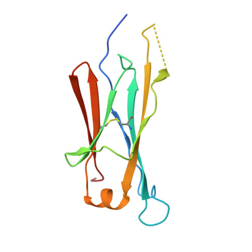
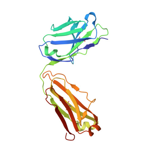
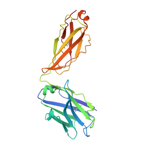
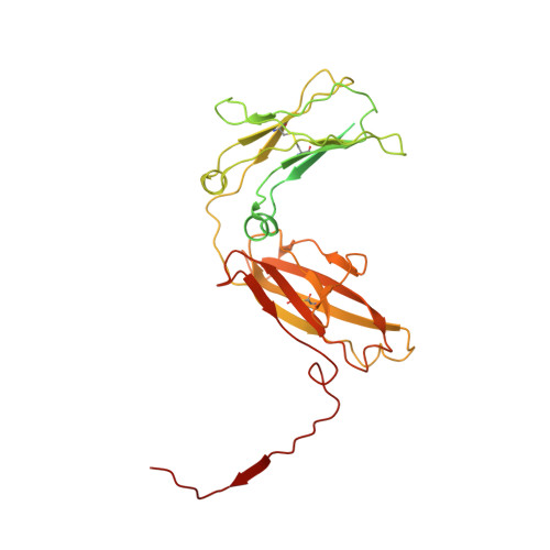
    Immunoglobulin M (IgM) is the most ancient of the five isotypes of immunoglobulin (Ig) molecules and serves as the first line of defence against pathogens. Here, we use cryo-EM to image the structure of the human full-length IgM pentamer, revealing antigen binding domains flexibly attached to the asymmetric and rigid core formed by the Cμ4 and Cμ3 constant regions and the J-chain. A hinge is located at the Cμ3/Cμ2 domain interface, allowing Fabs and Cμ2 to pivot as a unit both in-plane and out-of-plane. This motion is different from that observed in IgG and IgA, where the two Fab arms are able to swing independently. A biased orientation of one pair of Fab arms results from asymmetry in the constant domain (Cμ3) at the IgM subunit interacting most extensively with the J-chain. This may influence the multi-valent binding to surface-associated antigens and complement pathway activation. By comparison, the structure of the Fc fragment in the IgM monomer is similar to that of the pentamer, but is more dynamic in the Cμ4 domain.


  • Organizational Affiliation

    Structural Biology Science Technology Platform, The Francis Crick Institute, 1 Midland Road, London, NW1 1AT, UK.


Macromolecules
Find similar proteins by:  (by identity cutoff)  |  3D Structure
Entity ID: 1
MoleculeChains Sequence LengthOrganismDetailsImage
IgM C2-domain from mouse
A, B
117Homo sapiensMutation(s): 0 
Entity Groups  
Sequence Clusters30% Identity50% Identity70% Identity90% Identity95% Identity100% Identity
Sequence Annotations
Expand
  • Reference Sequence
Find similar proteins by:  (by identity cutoff)  |  3D Structure
Entity ID: 2
MoleculeChains Sequence LengthOrganismDetailsImage
IgM Fab, light chainC [auth L],
E [auth S]
215Homo sapiensMutation(s): 0 
Entity Groups  
Sequence Clusters30% Identity50% Identity70% Identity90% Identity95% Identity100% Identity
Sequence Annotations
Expand
  • Reference Sequence
Find similar proteins by:  (by identity cutoff)  |  3D Structure
Entity ID: 3
MoleculeChains Sequence LengthOrganismDetailsImage
IgM Fab, heavy chainD [auth H],
F [auth Q]
226Homo sapiensMutation(s): 0 
Entity Groups  
Sequence Clusters30% Identity50% Identity70% Identity90% Identity95% Identity100% Identity
Sequence Annotations
Expand
  • Reference Sequence
Find similar proteins by:  (by identity cutoff)  |  3D Structure
Entity ID: 4
MoleculeChains Sequence LengthOrganismDetailsImage
Immunoglobulin heavy constant mu383Homo sapiensMutation(s): 0 
UniProt & NIH Common Fund Data Resources
Find proteins for P01871 (Homo sapiens)
Explore P01871 
Go to UniProtKB:  P01871
PHAROS:  P01871
GTEx:  ENSG00000211899 
Entity Groups  
Sequence Clusters30% Identity50% Identity70% Identity90% Identity95% Identity100% Identity
UniProt GroupP01871
Sequence Annotations
Expand
  • Reference Sequence
Find similar proteins by:  (by identity cutoff)  |  3D Structure
Entity ID: 5
MoleculeChains Sequence LengthOrganismDetailsImage
Immunoglobulin J chainQ [auth J]136Homo sapiensMutation(s): 0 
UniProt & NIH Common Fund Data Resources
Find proteins for P01591 (Homo sapiens)
Explore P01591 
Go to UniProtKB:  P01591
PHAROS:  P01591
GTEx:  ENSG00000132465 
Entity Groups  
Sequence Clusters30% Identity50% Identity70% Identity90% Identity95% Identity100% Identity
UniProt GroupP01591
Sequence Annotations
Expand
  • Reference Sequence
Oligosaccharides

Help

Entity ID: 6
MoleculeChains Length2D Diagram Glycosylation3D Interactions
2-acetamido-2-deoxy-beta-D-glucopyranose-(1-4)-2-acetamido-2-deoxy-beta-D-glucopyranoseR [auth I],
S [auth M],
T [auth P]
2N-Glycosylation
Glycosylation Resources
GlyTouCan:  G42666HT
GlyCosmos:  G42666HT
GlyGen:  G42666HT
Experimental Data & Validation

Experimental Data

  • Method: ELECTRON MICROSCOPY
  • Resolution: 6.80 Å
  • Aggregation State: PARTICLE 
  • Reconstruction Method: SINGLE PARTICLE 

Structure Validation

View Full Validation Report


Entry History & Funding Information

Deposition Data


Funding OrganizationLocationGrant Number
Cancer Research UKUnited KingdomFC001143
Medical Research Council (MRC, United Kingdom)United KingdomFC001143
Wellcome TrustUnited KingdomFC001143
Cancer Research UKUnited KingdomFC001185
Medical Research Council (MRC, United Kingdom)United KingdomFC001185
Wellcome TrustUnited KingdomFC001185

Revision History  (Full details and data files)

  • Version 1.0: 2022-10-26
    Type: Initial release
  • Version 1.1: 2022-11-09
    Changes: Database references