8A2O | pdb_00008a2o

Room-temperature structure of the stabilised A2A-Theophylline complex determined by synchrotron serial crystallography


Experimental Data Snapshot

  • Method: X-RAY DIFFRACTION
  • Resolution: 3.45 Å
  • R-Value Free: 
    0.241 (Depositor), 0.240 (DCC) 
  • R-Value Work: 
    0.218 (Depositor), 0.218 (DCC) 
  • R-Value Observed: 
    0.220 (Depositor) 

Starting Model: experimental
View more details

wwPDB Validation 3D Report Full Report

Validation slider image for 8A2O

Ligand Structure Quality Assessment 


This is version 1.3 of the entry. See complete history

Literature

A versatile approach to high-density microcrystals in lipidic cubic phase for room-temperature serial crystallography.

Birch, J.Kwan, T.O.C.Judge, P.J.Axford, D.Aller, P.Butryn, A.Reis, R.I.Bada Juarez, J.F.Vinals, J.Owen, R.L.Nango, E.Tanaka, R.Tono, K.Joti, Y.Tanaka, T.Owada, S.Sugahara, M.Iwata, S.Orville, A.M.Watts, A.Moraes, I.

(2023) J Appl Crystallogr 56: 1361-1370

  • DOI: https://doi.org/10.1107/S1600576723006428
  • Primary Citation Related Structures: 
    7ZY3, 8A2O, 8A2P

  • PubMed Abstract: 

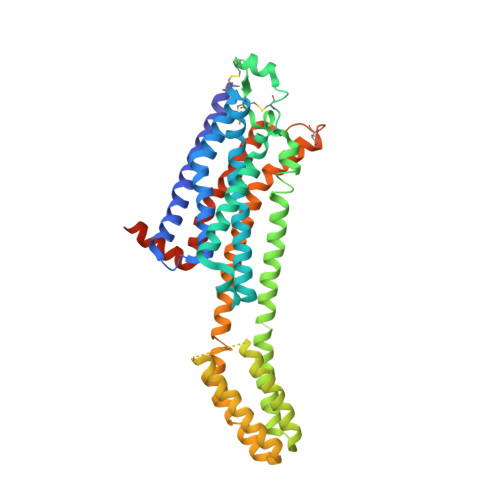
    Serial crystallography has emerged as an important tool for structural studies of integral membrane proteins. The ability to collect data from micrometre-sized weakly diffracting crystals at room temperature with minimal radiation damage has opened many new opportunities in time-resolved studies and drug discovery. However, the production of integral membrane protein microcrystals in lipidic cubic phase at the desired crystal density and quantity is challenging. This paper introduces VIALS (versatile approach to high-density microcrystals in lipidic cubic phase for serial crystallography), a simple, fast and efficient method for preparing hundreds of microlitres of high-density microcrystals suitable for serial X-ray diffraction experiments at both synchrotron and free-electron laser sources. The method is also of great benefit for rational structure-based drug design as it facilitates in situ crystal soaking and rapid determination of many co-crystal structures. Using the VIALS approach, room-temperature structures are reported of (i) the archaerhodopsin-3 protein in its dark-adapted state and 110 ns photocycle intermediate, determined to 2.2 and 1.7 Å, respectively, and (ii) the human A 2A adenosine receptor in complex with two different ligands determined to a resolution of 3.5 Å.


  • Organizational Affiliation
    • Membrane Protein Laboratory, Diamond Light Source, Harwell Science and Innovation Campus, Didcot, Oxfordshire OX11 0DE, United Kingdom.

Macromolecule Content 

  • Total Structure Weight: 49.85 kDa 
  • Atom Count: 3,188 
  • Modeled Residue Count: 386 
  • Deposited Residue Count: 423 
  • Unique protein chains: 1

Macromolecules

Find similar proteins by:|  3D Structure
Entity ID: 1
MoleculeChains  Sequence LengthOrganismDetailsImage
Adenosine receptor A2a,Soluble cytochrome b562423Homo sapiensMutation(s): 12 
Gene Names: ADORA2AADORA2cybC
Membrane Entity: Yes 
UniProt & NIH Common Fund Data Resources
Find proteins for P0ABE7 (Escherichia coli)
Explore P0ABE7 
Go to UniProtKB:  P0ABE7
Find proteins for P29274 (Homo sapiens)
Explore P29274 
Go to UniProtKB:  P29274
PHAROS:  P29274
GTEx:  ENSG00000128271 
Entity Groups
Sequence Clusters30% Identity50% Identity70% Identity90% Identity95% Identity100% Identity
UniProt GroupsP0ABE7P29274
Sequence Annotations
Expand
Reference Sequence

Small Molecules

Ligands 5 Unique
IDChains Name / Formula / InChI Key2D Diagram3D Interactions
CLR

Query on CLR


Download:Ideal Coordinates CCD File
C [auth A],
D [auth A],
E [auth A]
CHOLESTEROL
C27 H46 O
HVYWMOMLDIMFJA-DPAQBDIFSA-N
OLC

Query on OLC


Download:Ideal Coordinates CCD File
G [auth A],
H [auth A]
(2R)-2,3-dihydroxypropyl (9Z)-octadec-9-enoate
C21 H40 O4
RZRNAYUHWVFMIP-GDCKJWNLSA-N
OLA

Query on OLA


Download:Ideal Coordinates CCD File
F [auth A],
I [auth A],
J [auth A]
OLEIC ACID
C18 H34 O2
ZQPPMHVWECSIRJ-KTKRTIGZSA-N
TEP
(Subject of Investigation/LOI)

Query on TEP


Download:Ideal Coordinates CCD File
B [auth A]THEOPHYLLINE
C7 H8 N4 O2
ZFXYFBGIUFBOJW-UHFFFAOYSA-N
NA

Query on NA


Download:Ideal Coordinates CCD File
K [auth A]SODIUM ION
Na
FKNQFGJONOIPTF-UHFFFAOYSA-N

Experimental Data & Validation

Experimental Data

  • Method: X-RAY DIFFRACTION
  • Resolution: 3.45 Å
  • R-Value Free:  0.241 (Depositor), 0.240 (DCC) 
  • R-Value Work:  0.218 (Depositor), 0.218 (DCC) 
  • R-Value Observed: 0.220 (Depositor) 
Space Group: C 2 2 21
Unit Cell:
Length ( Å )Angle ( ˚ )
a = 40.531α = 90
b = 182.311β = 90
c = 144.267γ = 90
Software Package:
Software NamePurpose
REFMACrefinement
PDB_EXTRACTdata extraction
DIALSdata reduction
PHASERphasing

Structure Validation

View Full Validation Report


Ligand Structure Quality Assessment 


Entry History 

& Funding Information

Deposition Data


Funding OrganizationLocationGrant Number
Other governmentUnited Kingdom--

Revision History  (Full details and data files)

  • Version 1.0: 2023-08-30
    Type: Initial release
  • Version 1.1: 2023-10-18
    Changes: Database references
  • Version 1.2: 2024-10-09
    Changes: Structure summary
  • Version 1.3: 2026-03-04
    Changes: Refinement description