8A1E

Rabies virus glycoprotein in complex with Fab fragments of 17C7 and 1112-1 neutralizing antibodies


Experimental Data Snapshot

  • Method: ELECTRON MICROSCOPY
  • Resolution: 2.83 Å
  • Aggregation State: PARTICLE 
  • Reconstruction Method: SINGLE PARTICLE 

wwPDB Validation   3D Report Full Report


This is version 1.2 of the entry. See complete history


Literature

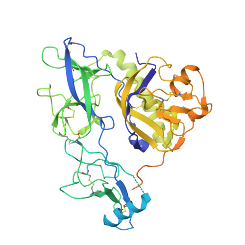
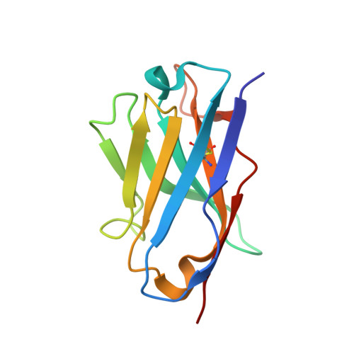
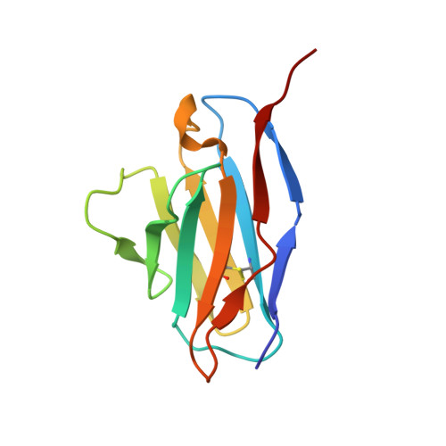
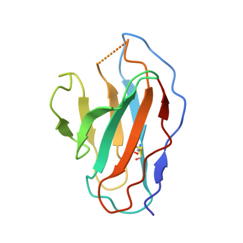
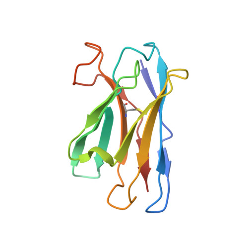
Structure of trimeric pre-fusion rabies virus glycoprotein in complex with two protective antibodies.

Ng, W.M.Fedosyuk, S.English, S.Augusto, G.Berg, A.Thorley, L.Haselon, A.S.Segireddy, R.R.Bowden, T.A.Douglas, A.D.

(2022) Cell Host Microbe 30: 1219-1230.e7

  • DOI: https://doi.org/10.1016/j.chom.2022.07.014
  • Primary Citation of Related Structures:  
    8A1E

  • PubMed Abstract: 

    Rabies virus (RABV) causes lethal encephalitis and is responsible for approximately 60,000 deaths per year. As the sole virion-surface protein, the rabies virus glycoprotein (RABV-G) mediates host-cell entry. RABV-G's pre-fusion trimeric conformation displays epitopes bound by protective neutralizing antibodies that can be induced by vaccination or passively administered for post-exposure prophylaxis. We report a 2.8-Å structure of a RABV-G trimer in the pre-fusion conformation, in complex with two neutralizing and protective monoclonal antibodies, 17C7 and 1112-1, that recognize distinct epitopes. One of these antibodies is a licensed prophylactic (17C7, Rabishield), which we show locks the protein in pre-fusion conformation. Targeted mutations can similarly stabilize RABV-G in the pre-fusion conformation, a key step toward structure-guided vaccine design. These data reveal the higher-order architecture of a key therapeutic target and the structural basis of neutralization by antibodies binding two key antigenic sites, and this will facilitate the development of improved vaccines and prophylactic antibodies.


  • Organizational Affiliation

    Jenner Institute, Old Road Campus Research Building, Roosevelt Drive, Oxford OX3 7DQ, UK; Division of Structural Biology, Wellcome Centre for Human Genetics, University of Oxford, Roosevelt Drive, Oxford OX3 7BN, UK.


Macromolecules
Find similar proteins by:  (by identity cutoff)  |  3D Structure
Entity ID: 1
MoleculeChains Sequence LengthOrganismDetailsImage
Glycoprotein505Rabies virus strain Pasteur vaccinMutation(s): 0 
UniProt
Find proteins for P08667 (Rabies virus (strain Pasteur vaccins / PV))
Explore P08667 
Go to UniProtKB:  P08667
Entity Groups  
Sequence Clusters30% Identity50% Identity70% Identity90% Identity95% Identity100% Identity
UniProt GroupP08667
Sequence Annotations
Expand
  • Reference Sequence
Find similar proteins by:  (by identity cutoff)  |  3D Structure
Entity ID: 2
MoleculeChains Sequence LengthOrganismDetailsImage
Fab 17C7 heavy chain variable domainB [auth D]119Homo sapiensMutation(s): 0 
Entity Groups  
Sequence Clusters30% Identity50% Identity70% Identity90% Identity95% Identity100% Identity
Sequence Annotations
Expand
  • Reference Sequence
Find similar proteins by:  (by identity cutoff)  |  3D Structure
Entity ID: 3
MoleculeChains Sequence LengthOrganismDetailsImage
Fab 17C7 light chain variable domainC [auth E]109Homo sapiensMutation(s): 0 
Entity Groups  
Sequence Clusters30% Identity50% Identity70% Identity90% Identity95% Identity100% Identity
Sequence Annotations
Expand
  • Reference Sequence
Find similar proteins by:  (by identity cutoff)  |  3D Structure
Entity ID: 4
MoleculeChains Sequence LengthOrganismDetailsImage
Fab 1112-1 light chain variable domainD [auth C]107Mus musculusMutation(s): 0 
Entity Groups  
Sequence Clusters30% Identity50% Identity70% Identity90% Identity95% Identity100% Identity
Sequence Annotations
Expand
  • Reference Sequence
Find similar proteins by:  (by identity cutoff)  |  3D Structure
Entity ID: 5
MoleculeChains Sequence LengthOrganismDetailsImage
Fab 1112-1 heavy chain variable domainE [auth B]121Mus musculusMutation(s): 0 
Entity Groups  
Sequence Clusters30% Identity50% Identity70% Identity90% Identity95% Identity100% Identity
Sequence Annotations
Expand
  • Reference Sequence
Experimental Data & Validation

Experimental Data

  • Method: ELECTRON MICROSCOPY
  • Resolution: 2.83 Å
  • Aggregation State: PARTICLE 
  • Reconstruction Method: SINGLE PARTICLE 
EM Software:
TaskSoftware PackageVersion
RECONSTRUCTIONcryoSPARC3.1
MODEL REFINEMENTPHENIX1.19.2_4158

Structure Validation

View Full Validation Report


Entry History & Funding Information

Deposition Data


Funding OrganizationLocationGrant Number
Wellcome TrustUnited Kingdom220679/Z/20/Z
Medical Research Council (MRC, United Kingdom)United KingdomMR/P017339/1
Medical Research Council (MRC, United Kingdom)United KingdomMR/S007555/1
Marie Sklodowska-Curie Actions, FragNET ITNEuropean Union840866
Wellcome TrustUnited Kingdom060208/Z/00/Z
Wellcome TrustUnited Kingdom093305/Z/10/Z
Wellcome TrustUnited Kingdom203141/Z/16/Z

Revision History  (Full details and data files)

  • Version 1.0: 2022-08-17
    Type: Initial release
  • Version 1.1: 2022-08-31
    Changes: Database references
  • Version 1.2: 2022-09-28
    Changes: Database references