8ZWQ | pdb_00008zwq

pseudorabies virus dUTPase structure


Experimental Data Snapshot

  • Method: X-RAY DIFFRACTION
  • Resolution: 2.23 Å
  • R-Value Free: 
    0.339 (Depositor), 0.337 (DCC) 
  • R-Value Work: 
    0.277 (Depositor), 0.277 (DCC) 
  • R-Value Observed: 
    0.280 (Depositor) 

Starting Model: in silico
View more details

wwPDB Validation   3D Report Full Report


Ligand Structure Quality Assessment 


This is version 1.1 of the entry. See complete history


Literature

Structural definition of pseudorabies virus dUTPase reveals a novel folding dimer in the herpesvirus family.

Li, J.Ma, S.Yang, R.Xu, J.Wang, Y.Ye, S.

(2024) Int J Biol Macromol 280: 135696-135696

  • DOI: https://doi.org/10.1016/j.ijbiomac.2024.135696
  • Primary Citation of Related Structures:  
    8ZWQ

  • PubMed Abstract: 

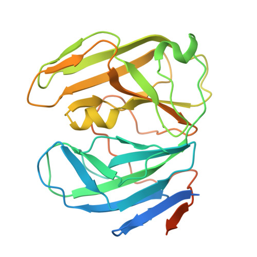
    The pseudorabies virus (PRV) causes severe and fatal acute respiratory disease in pigs. During PRV proliferation, the enzyme deoxyuridine 5'-triphosphate nucleotide hydrolase (dUTPase) plays a pivotal role in maintaining a low dUTP/dTTP ratio, thereby ensuring the accuracy of viral DNA replication. However, its structure and catalytic mechanisms have not been fully elucidated. Here, we report the crystal structure of PRV dUTPase at a 2.24 Å resolution and demonstrate an unprecedented dimeric architecture, with a conserved enzyme activity center of the herpesvirus family. The enzyme activity center is located in a cavity between the two domains, forming a pocket for binding substrate dUMP and magnesium ions. Remarkably, the exquisite interface of the dimer is primarily composed of four antiparallel β-sheets, which form 11 hydrogen bonds between the residues P33-V36 and R242-A248 to maintain protein stability. To the best of our knowledge, this is the first report demonstrating that dUTPase exists as a dimer in the herpesvirus family. These findings not only present a novel fold dimeric structure but also deepen the scope of our comprehension of structural diversity in dUTPase family.


  • Organizational Affiliation
    • Frontiers Science Center for Synthetic Biology (Ministry of Education), Tianjin Key Laboratory of Function and Application of Biological Macromolecular Structures, School of Life Sciences, Tianjin University, Tianjin, China.

Macromolecules
Find similar proteins by:  (by identity cutoff)  |  3D Structure
Entity ID: 1
MoleculeChains Sequence LengthOrganismDetailsImage
dUTPaseA,
B [auth C],
C [auth B],
D
276Suid alphaherpesvirus 1Mutation(s): 0 
Gene Names: UL50
EC: 3.6.1.23
UniProt
Find proteins for Q90030 (Suid herpesvirus 1 (strain Kaplan))
Explore Q90030 
Go to UniProtKB:  Q90030
Entity Groups  
Sequence Clusters30% Identity50% Identity70% Identity90% Identity95% Identity100% Identity
UniProt GroupQ90030
Sequence Annotations
Expand
  • Reference Sequence
Experimental Data & Validation

Experimental Data

  • Method: X-RAY DIFFRACTION
  • Resolution: 2.23 Å
  • R-Value Free:  0.339 (Depositor), 0.337 (DCC) 
  • R-Value Work:  0.277 (Depositor), 0.277 (DCC) 
  • R-Value Observed: 0.280 (Depositor) 
Space Group: C 1 2 1
Unit Cell:
Length ( Å )Angle ( ˚ )
a = 98.249α = 90
b = 103.264β = 99.472
c = 98.733γ = 90
Software Package:
Software NamePurpose
PHENIXrefinement
HKL-3000data reduction
HKL-3000data scaling
PHASERphasing

Structure Validation

View Full Validation Report


Ligand Structure Quality Assessment 


Entry History & Funding Information

Deposition Data

  • Released Date: 2025-06-18 
  • Deposition Author(s): Wang, Y.

Funding OrganizationLocationGrant Number
National Natural Science Foundation of China (NSFC)China81801998

Revision History  (Full details and data files)

  • Version 1.0: 2025-06-18
    Type: Initial release
  • Version 1.1: 2025-12-31
    Changes: Database references