The characterization and structural basis of a human broadly binding antibody to HBV core protein.
Yan, H., Liu, C., Li, Y., Tang, S., Guo, H., Zhou, B., Fan, Q., Wang, H., Ge, X., Wang, X., Liao, X., Li, J., Zhang, Z., Ju, B.(2025) J Virol 99: e0169424-e0169424
- PubMed: 39601613 Search on PubMed
- DOI: https://doi.org/10.1128/jvi.01694-24
- Primary Citation Related Structures:
8ZRE, 8ZRH, 8ZRR - PubMed Abstract:
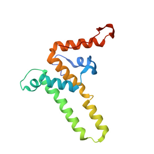
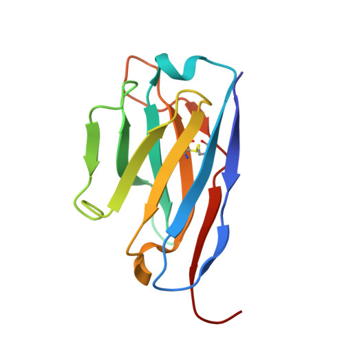
Hepatitis B virus (HBV) core protein (HBc) plays a crucial role in the virus life cycle, making it an important detection marker for HBV infection and a potential target for treatment. However, several commercially available monoclonal antibodies (mAbs) or polyclonal antibodies (pAbs) targeting HBc have certain limitations in detecting HBV across different genotypes in various biochemical assays, such as enzyme-linked immunosorbent assay, western blot, immunofluorescence assay, flow cytometry, and immune spot assay. In this study, we identified 12 human anti-HBc mAbs and evaluated their potential application in multiple biochemical assays. These mAbs mainly recognized the epitopes near residues 20-22 amino acids (a.a.) and 77-78 a.a. of HBc, demonstrating a broadly cross-genotypic activity. Notably, three Group II mAbs, named cAbA1, cAbD4, and cAbF9, displayed excellent capacities for the detection of HBV infection in all tested biochemical assays. Furthermore, we determined a 3.22 Å of cryo-electron microscopy (cryo-EM) structure of the fragment of antigen binding (Fab) of cAbD4 complexed with HBc dimer, which was the highest resolution of the structural model for Fab-HBc to date. Collectively, our findings provided excellent and reliable antibody candidates for live HBV detection and revealed the recognition mechanism and the detailed interaction information of a potent human anti-HBc mAb binding to the spike tips of HBc dimer.IMPORTANCEThe lack of excellent detection Abs for live hepatitis B virus (HBV) infection and high-resolution structures of the Ab-HBV core protein (HBc) complex largely limited the development of HBV-related research. This study reports a panel of anti-HBc monoclonal antibodies (mAbs) with excellent capacities for detecting HBV infection in multiple biochemical assays and determines a 3.22 Å of cryo-EM structure of HBc with a potent binding mAb. These findings provide excellent and reliable detecting tools for HBV-related research and promote the understanding of the recognition mechanism of anti-HBc mAbs to HBc particles.
- Institute for Hepatology, National Clinical Research Center for Infectious Disease, Shenzhen Third People's Hospital, Shenzhen, Guangdong Province, China.
Organizational Affiliation: