


preF8P of RSV glycoprotein F0
Huang, Q., Lang, Q., Han, X., Yan, J.To be published.
Experimental Data Snapshot
Starting Model: in silico
View more details
wwPDB Validation 3D Report Full Report

Entity ID: 1 | |||||
|---|---|---|---|---|---|
| Molecule | Chains | Sequence Length | Organism | Details | Image |
| Fusion glycoprotein F0,Fibritin | 522 | human respiratory syncytial virus, Enterobacteria phage T6 | Mutation(s): 0 | 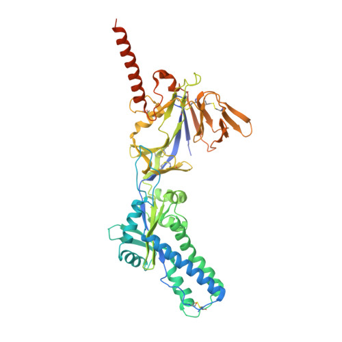 | |
UniProt | |||||
Find proteins for A0A346FJN8 (Enterobacteria phage T6) Explore A0A346FJN8 Go to UniProtKB: A0A346FJN8 | |||||
Entity Groups | |||||
| Sequence Clusters | 30% Identity50% Identity70% Identity90% Identity95% Identity100% Identity | ||||
| UniProt Groups | P12568A0A346FJN8 | ||||
Sequence AnnotationsExpand | |||||
Reference Sequence | |||||
| Length ( Å ) | Angle ( ˚ ) |
|---|---|
| a = 158.423 | α = 90 |
| b = 168.696 | β = 90 |
| c = 190.683 | γ = 90 |
| Software Name | Purpose |
|---|---|
| PHENIX | refinement |
| XDS | data scaling |
| XDS | data reduction |
| PHASER | phasing |
| Funding Organization | Location | Grant Number |
|---|---|---|
| National Natural Science Foundation of China (NSFC) | China | -- |


