8ZPW | pdb_00008zpw

Cryo-EM structure of the yeast Htm1/Pdi1 complex at a resolution of 3.0 angstrom


Experimental Data Snapshot

  • Method: ELECTRON MICROSCOPY
  • Resolution: 3.00 Å
  • Aggregation State: PARTICLE 
  • Reconstruction Method: SINGLE PARTICLE 

wwPDB Validation   3D Report Full Report


This is version 1.2 of the entry. See complete history


Literature

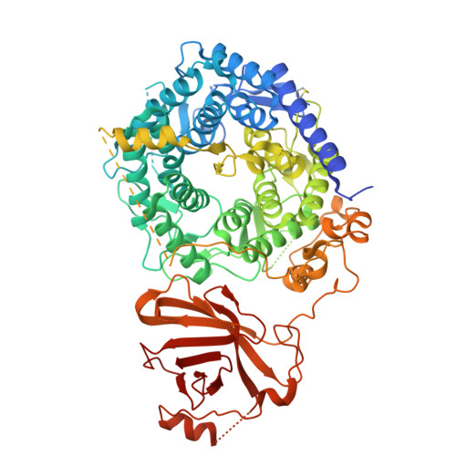
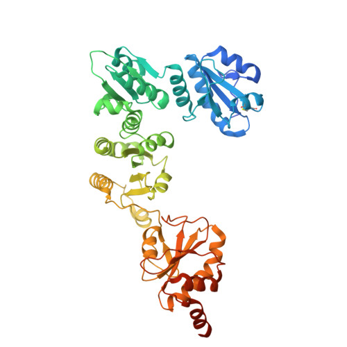
Initiation of ERAD by the bifunctional complex of Mnl1 mannosidase and protein disulfide isomerase.

Zhao, D.Wu, X.Rapoport, T.A.

(2024) bioRxiv 

  • DOI: https://doi.org/10.1101/2024.10.17.618908
  • Primary Citation of Related Structures:  
    8ZPW

  • PubMed Abstract: 

    Misfolded glycoproteins in the endoplasmic reticulum (ER) lumen are translocated into the cytosol and degraded by the proteasome, a conserved process called ER-associated protein degradation (ERAD). In S. cerevisiae , the glycan of these proteins is trimmed by the luminal mannosidase Mnl1 (Htm1) to generate a signal that triggers degradation. Curiously, Mnl1 is permanently associated with protein disulfide isomerase (Pdi1). Here, we have used cryo- electron microscopy, biochemical, and in vivo experiments to clarify how this complex initiates ERAD. The Mnl1-Pdi1 complex first de-mannosylates misfolded, globular proteins that are recognized through a C-terminal domain (CTD) of Mnl1; Pdi1 causes the CTD to ignore completely unfolded polypeptides. The disulfides of these globular proteins are then reduced by the Pdi1 component of the complex, generating unfolded polypeptides that can be translocated across the membrane. Mnl1 blocks the canonical oxidative function of Pdi1, but allows it to function as the elusive disulfide reductase in ERAD.


Macromolecules
Find similar proteins by:  (by identity cutoff)  |  3D Structure
Entity ID: 1
MoleculeChains Sequence LengthOrganismDetailsImage
ER degradation-enhancing alpha-mannosidase-like protein 1796Saccharomyces cerevisiaeMutation(s): 0 
Gene Names: MNL1HTM1YHR204W
EC: 3.2.1.24
UniProt
Find proteins for P38888 (Saccharomyces cerevisiae (strain ATCC 204508 / S288c))
Explore P38888 
Go to UniProtKB:  P38888
Entity Groups  
Sequence Clusters30% Identity50% Identity70% Identity90% Identity95% Identity100% Identity
UniProt GroupP38888
Sequence Annotations
Expand
  • Reference Sequence
Find similar proteins by:  (by identity cutoff)  |  3D Structure
Entity ID: 2
MoleculeChains Sequence LengthOrganismDetailsImage
Protein disulfide-isomerase522Saccharomyces cerevisiaeMutation(s): 0 
Gene Names: PDI1MFP1TRG1YCL043CYCL313YCL43C
EC: 5.3.4.1
UniProt
Find proteins for P17967 (Saccharomyces cerevisiae (strain ATCC 204508 / S288c))
Explore P17967 
Go to UniProtKB:  P17967
Entity Groups  
Sequence Clusters30% Identity50% Identity70% Identity90% Identity95% Identity100% Identity
UniProt GroupP17967
Sequence Annotations
Expand
  • Reference Sequence
Experimental Data & Validation

Experimental Data

  • Method: ELECTRON MICROSCOPY
  • Resolution: 3.00 Å
  • Aggregation State: PARTICLE 
  • Reconstruction Method: SINGLE PARTICLE 
EM Software:
TaskSoftware PackageVersion
MODEL REFINEMENTPHENIX

Structure Validation

View Full Validation Report


Entry History & Funding Information

Deposition Data


Funding OrganizationLocationGrant Number
National Institutes of Health/National Institute of General Medical Sciences (NIH/NIGMS)United StatesR01 GM052586

Revision History  (Full details and data files)

  • Version 1.0: 2024-11-13
    Type: Initial release
  • Version 1.1: 2024-11-27
    Changes: Data collection
  • Version 1.2: 2025-07-02
    Changes: Data collection