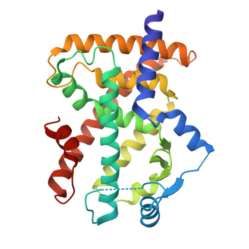
8ZNN | pdb_00008znn

X-ray structure of human PPAR delta ligand binding domain in complex with a synthetic agonist 16a


Experimental Data Snapshot

  • Method: X-RAY DIFFRACTION
  • Resolution: 1.95 Å
  • R-Value Free: 
    0.227 (Depositor), 0.228 (DCC) 
  • R-Value Work: 
    0.190 (Depositor), 0.191 (DCC) 

wwPDB Validation 3D Report Full Report

Validation slider image for 8ZNN

Ligand Structure Quality Assessment 


This is version 1.1 of the entry. See complete history

Literature

Design, Synthesis, and Biological Evaluation of Arylimidazole Derivatives as Potent PPAR delta Agonists for the Treatment of Renal Fibrosis.

Feng, Z.Xie, J.Hou, D.Fu, Z.Sun, S.Liu, X.Sun, G.Zheng, R.Liu, L.Xu, Q.Wen, X.Zhang, D.Yuan, H.Sun, H.Dai, L.

(2026) J Med Chem 69: 4469-4492

  • DOI: https://doi.org/10.1021/acs.jmedchem.5c03132
  • Primary Citation Related Structures: 
    8ZNN

  • PubMed Abstract: 

    Peroxisome proliferator-activator receptor δ (PPARδ) is ubiquitously expressed in the kidney, and its agonists are increasingly being recognized as a potential therapeutic strategy for renal diseases. In this work, we developed a series of arylimidazole derivatives as potent PPARδ agonists. Among them, compound 16a exhibited potent PPARδ agonistic activity (EC 50 = 0.50 nM) and high selectivity over PPARα/γ and some other nuclear receptors. The X-ray cocrystal structure revealed the binding mode of 16a and PPARδ at 1.94 Å resolution. Remarkably, compound 16a exhibited acceptable pharmacokinetic properties and good safety profiles in vivo and showed antirenal fibrosis effects in a dose-dependent manner in a mouse model of unilateral ureteral obstruction. Mechanistically, 16a activated PPARδ to restore fatty acid oxidation to attenuate TGF-β1-induced renal fibroblast activation. Collectively, 16a warrants further investigation as a promising drug candidate for treating renal fibrosis.


  • Organizational Affiliation
    • Jiangsu Key Laboratory of Drug Discovery for Metabolic Disease, State Key Laboratory of Natural Medicines, China Pharmaceutical University, Nanjing 210009, China.

Macromolecule Content 

  • Total Structure Weight: 31.39 kDa 
  • Atom Count: 2,386 
  • Modeled Residue Count: 260 
  • Deposited Residue Count: 270 
  • Unique protein chains: 1

Macromolecules

Find similar proteins by:|  3D Structure
Entity ID: 1
MoleculeChains  Sequence LengthOrganismDetailsImage
Peroxisome proliferator-activated receptor delta270Homo sapiensMutation(s): 0 
Gene Names: PPARDNR1C2PPARB
UniProt & NIH Common Fund Data Resources
Find proteins for Q03181 (Homo sapiens)
Explore Q03181 
Go to UniProtKB:  Q03181
PHAROS:  Q03181
GTEx:  ENSG00000112033 
Entity Groups
Sequence Clusters30% Identity50% Identity70% Identity90% Identity95% Identity100% Identity
UniProt GroupQ03181
Sequence Annotations
Expand
Reference Sequence

Small Molecules

Ligands 1 Unique
IDChains Name / Formula / InChI Key2D Diagram3D Interactions
A1D86
(Subject of Investigation/LOI)

Query on A1D86


Download:Ideal Coordinates CCD File
B [auth A]6-[2-[[2-[4-(trifluoromethyloxy)phenyl]benzimidazol-1-yl]methyl]phenoxy]hexanoic acid
C27 H25 F3 N2 O4
CIJJQZAOSIHXTJ-UHFFFAOYSA-N

Experimental Data & Validation

Experimental Data

  • Method: X-RAY DIFFRACTION
  • Resolution: 1.95 Å
  • R-Value Free:  0.227 (Depositor), 0.228 (DCC) 
  • R-Value Work:  0.190 (Depositor), 0.191 (DCC) 
Space Group: P 2 21 21
Unit Cell:
Length ( Å )Angle ( ˚ )
a = 37.228α = 90
b = 93.553β = 90
c = 95.962γ = 90
Software Package:
Software NamePurpose
PHENIXrefinement
PDB_EXTRACTdata extraction
HKL-2000data reduction
HKL-2000data scaling
PHENIXphasing

Structure Validation

View Full Validation Report


Ligand Structure Quality Assessment 


Entry History 

& Funding Information

Deposition Data


Funding OrganizationLocationGrant Number
Not fundedChina--

Revision History  (Full details and data files)

  • Version 1.0: 2025-12-24
    Type: Initial release
  • Version 1.1: 2026-07-08
    Changes: Database references