Evolutionary origins of self-sustained Kai protein circadian oscillators in cyanobacteria.
Mukaiyama, A., Furuike, Y., Ito-Miwa, K., Onoue, Y., Horiuchi, K., Kondo, K., Yamashita, E., Akiyama, S.(2025) Nat Commun 16: 4541-4541
- PubMed: 40374681 Search on PubMedSearch on PubMed Central
- DOI: https://doi.org/10.1038/s41467-025-59908-7
- Primary Citation Related Structures:
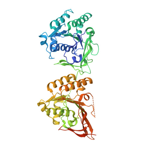
8ZN5, 8ZN6, 8ZN7 - PubMed Abstract:
Light-dark cycles affect photosynthetic efficiency in autotrophic cyanobacteria; therefore, determining whether ancient cyanobacteria possessed a self-sustained circadian clock when oxygenic photosynthetic systems were established is an important issue in chronobiology. Here we examine the oscillation of the clock protein KaiC in modern cyanobacteria, as well as the function and structure of ancestral Kai proteins, to determine the evolutionary origin of the self-sustained Kai-protein oscillators. The results show that the oldest double-domain KaiC in ancestral bacteria lacks the factors functionally and structurally essential for rhythmicity. The ancestral Kai proteins have acquired these factors through molecular evolution that occurred around Global Oxidation and Snowball Earth events, and are eventually inherited as a self-sustained circadian oscillator by the most recent common ancestor of cyanobacteria capable of oxygenic photosynthesis. This autonomous Kai protein oscillator is further inherited by most freshwater and marine cyanobacteria present today as an autotrophic basis for time-optimal acquisition and consumption of energy from oxygenic photosynthesis.
- Department of Bioscience and Biotechnology, Fukui Prefectural University, Eiheiji, 910-1195, Japan. amukai@fpu.ac.jp.
Organizational Affiliation: