8ZGG | pdb_00008zgg

Human lysine O-link glycosylation complex, LH3/ColGalT1 with bound UDP-glucose


Experimental Data Snapshot

  • Method: ELECTRON MICROSCOPY
  • Resolution: 3.75 Å
  • Aggregation State: PARTICLE 
  • Reconstruction Method: SINGLE PARTICLE 

wwPDB Validation   3D Report Full Report


This is version 1.1 of the entry. See complete history


Literature

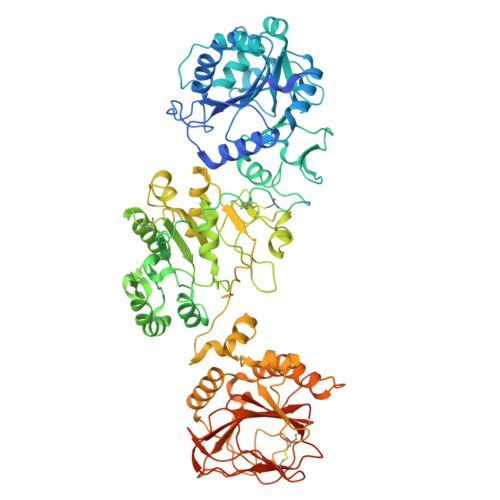
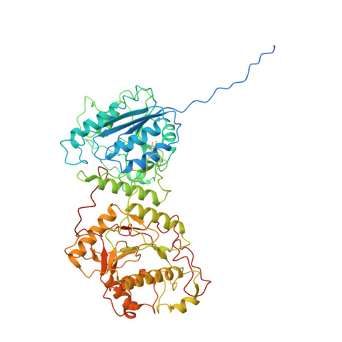
The structural basis for the human procollagen lysine hydroxylation and dual-glycosylation.

Peng, J.Li, W.Yao, D.Xia, Y.Wang, Q.Cai, Y.Li, S.Cao, M.Shen, Y.Ma, P.Liao, R.Zhao, J.Qin, A.Cao, Y.

(2025) Nat Commun 16: 2436-2436

  • DOI: https://doi.org/10.1038/s41467-025-57768-9
  • Primary Citation of Related Structures:  
    8ZGC, 8ZGE, 8ZGG, 8ZGH

  • PubMed Abstract: 

    The proper assembly and maturation of collagens necessitate the orchestrated hydroxylation and glycosylation of multiple lysyl residues in procollagen chains. Dysfunctions in this multistep modification process can lead to severe collagen-associated diseases. To elucidate the coordination of lysyl processing activities, we determine the cryo-EM structures of the enzyme complex formed by LH3/PLOD3 and GLT25D1/ColGalT1, designated as the KOGG complex. Our structural analysis reveals a tetrameric complex comprising dimeric LH3/PLOD3s and GLT25D1/ColGalT1s, assembled with interactions involving the N-terminal loop of GLT25D1/ColGalT1 bridging another GLT25D1/ColGalT1 and LH3/PLOD3. We further elucidate the spatial configuration of the hydroxylase, galactosyltransferase, and glucosyltransferase sites within the KOGG complex, along with the key residues involved in substrate binding at these enzymatic sites. Intriguingly, we identify a high-order oligomeric pattern characterized by the formation of a fiber-like KOGG polymer assembled through the repetitive incorporation of KOGG tetramers as the biological unit.


  • Organizational Affiliation
    • Department of Orthopaedics, Shanghai Key Laboratory of Orthopaedic Implant, Shanghai Ninth People's Hospital, Shanghai Jiao Tong University School of Medicine, Shanghai, 200011, China.

Macromolecules
Find similar proteins by:  (by identity cutoff)  |  3D Structure
Entity ID: 1
MoleculeChains Sequence LengthOrganismDetailsImage
Multifunctional procollagen lysine hydroxylase and glycosyltransferase LH3
A, B, C, D
778Homo sapiensMutation(s): 0 
Gene Names: PLOD3
EC: 1.14.11.4 (PDB Primary Data), 2.4.1.50 (PDB Primary Data), 2.4.1.66 (PDB Primary Data)
UniProt & NIH Common Fund Data Resources
Find proteins for O60568 (Homo sapiens)
Explore O60568 
Go to UniProtKB:  O60568
PHAROS:  O60568
GTEx:  ENSG00000106397 
Entity Groups  
Sequence Clusters30% Identity50% Identity70% Identity90% Identity95% Identity100% Identity
UniProt GroupO60568
Glycosylation
Glycosylation Sites: 2Go to GlyGen: O60568-1
Sequence Annotations
Expand
  • Reference Sequence
Find similar proteins by:  (by identity cutoff)  |  3D Structure
Entity ID: 2
MoleculeChains Sequence LengthOrganismDetailsImage
Procollagen galactosyltransferase 1E [auth U],
F [auth V],
G [auth W],
H [auth X]
653Homo sapiensMutation(s): 0 
Gene Names: COLGALT1GLT25D1PSEC0241
EC: 2.4.1.50
UniProt & NIH Common Fund Data Resources
Find proteins for Q8NBJ5 (Homo sapiens)
Explore Q8NBJ5 
Go to UniProtKB:  Q8NBJ5
PHAROS:  Q8NBJ5
GTEx:  ENSG00000130309 
Entity Groups  
Sequence Clusters30% Identity50% Identity70% Identity90% Identity95% Identity100% Identity
UniProt GroupQ8NBJ5
Glycosylation
Glycosylation Sites: 1Go to GlyGen: Q8NBJ5-1
Sequence Annotations
Expand
  • Reference Sequence
Oligosaccharides

Help

Entity ID: 3
MoleculeChains Length2D Diagram Glycosylation3D Interactions
2-acetamido-2-deoxy-beta-D-glucopyranose-(1-4)-2-acetamido-2-deoxy-beta-D-glucopyranoseI [auth E],
J [auth F]
2N-Glycosylation
Glycosylation Resources
GlyTouCan:  G42666HT
GlyCosmos:  G42666HT
GlyGen:  G42666HT
Small Molecules
Ligands 6 Unique
IDChains Name / Formula / InChI Key2D Diagram3D Interactions
660 (Subject of Investigation/LOI)
Query on 660

Download Ideal Coordinates CCD File 
V [auth U],
X [auth V]
[[(2~{R},3~{S},4~{R},5~{R})-5-[2,4-bis(oxidanylidene)pyrimidin-1-yl]-3,4-bis(oxidanyl)oxolan-2-yl]methoxy-oxidanyl-phosphoryl]oxy-[[(2~{S},3~{R},4~{S},5~{S},6~{R})-6-(hydroxymethyl)-3,4,5-tris(oxidanyl)oxan-2-yl]methyl]phosphinic acid
C16 H26 N2 O16 P2
WUPLBUVQIJIOHV-RIDHHKDKSA-N
UDP (Subject of Investigation/LOI)
Query on UDP

Download Ideal Coordinates CCD File 
N [auth A],
T [auth B]
URIDINE-5'-DIPHOSPHATE
C9 H14 N2 O12 P2
XCCTYIAWTASOJW-XVFCMESISA-N
NAG (Subject of Investigation/LOI)
Query on NAG

Download Ideal Coordinates CCD File 
K [auth A],
P [auth B],
Q [auth B]
2-acetamido-2-deoxy-beta-D-glucopyranose
C8 H15 N O6
OVRNDRQMDRJTHS-FMDGEEDCSA-N
AKG (Subject of Investigation/LOI)
Query on AKG

Download Ideal Coordinates CCD File 
L [auth A],
R [auth B]
2-OXOGLUTARIC ACID
C5 H6 O5
KPGXRSRHYNQIFN-UHFFFAOYSA-N
FE2 (Subject of Investigation/LOI)
Query on FE2

Download Ideal Coordinates CCD File 
M [auth A],
S [auth B]
FE (II) ION
Fe
CWYNVVGOOAEACU-UHFFFAOYSA-N
MN (Subject of Investigation/LOI)
Query on MN

Download Ideal Coordinates CCD File 
O [auth A],
U [auth B],
W [auth U],
Y [auth V]
MANGANESE (II) ION
Mn
WAEMQWOKJMHJLA-UHFFFAOYSA-N
Experimental Data & Validation

Experimental Data

  • Method: ELECTRON MICROSCOPY
  • Resolution: 3.75 Å
  • Aggregation State: PARTICLE 
  • Reconstruction Method: SINGLE PARTICLE 
EM Software:
TaskSoftware PackageVersion
MODEL REFINEMENTPHENIX1.20.1_4487:

Structure Validation

View Full Validation Report


Entry History & Funding Information

Deposition Data


Funding OrganizationLocationGrant Number
National Natural Science Foundation of China (NSFC)China82072468
National Natural Science Foundation of China (NSFC)China82272519

Revision History  (Full details and data files)

  • Version 1.0: 2025-03-19
    Type: Initial release
  • Version 1.1: 2025-03-26
    Changes: Data collection, Database references