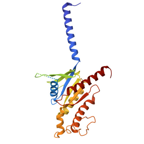
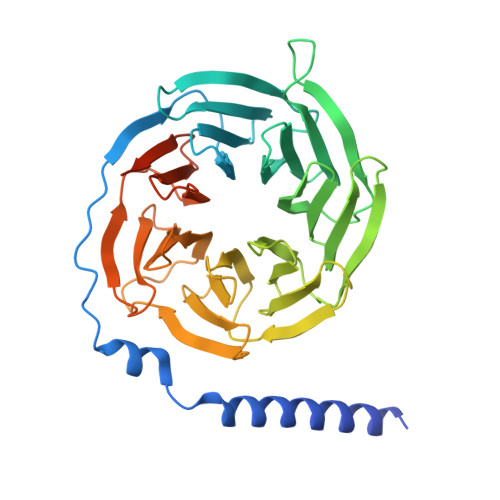
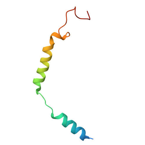
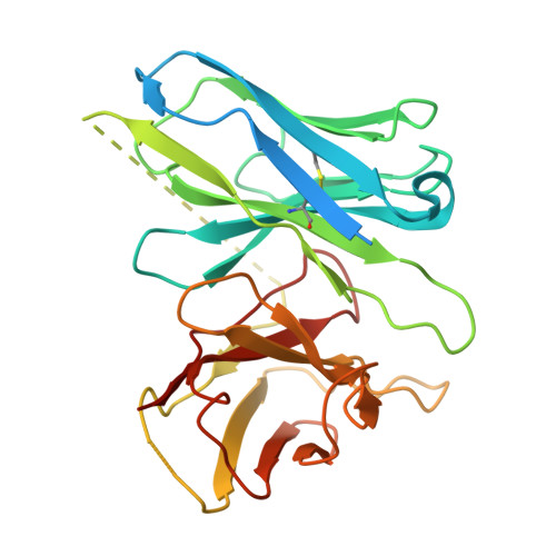
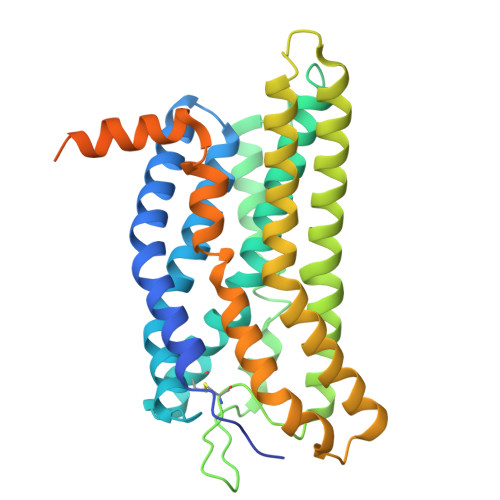
Animals have evolved pH-sensing membrane receptors, such as G-protein-coupled receptor 4 (GPR4), to monitor pH changes related to their physiology and generate adaptive reactions. However, the evolutionary trajectory and structural mechanism of proton sensing by GPR4 remain unresolved. Here, we observed a positive correlation between the optimal pH of GPR4 activity and the blood pH range across different species. By solving 7-cryoelectron microscopy (cryo-EM) structures of Xenopus tropicalis GPR4 (xtGPR4) and Mus musculus GPR4 (mmGPR4) under varying pH conditions, we identified that protonation of H ECL2-45.47 and H 7.36 enabled polar network establishment and tighter association between the extracellular loop 2 (ECL2) and 7 transmembrane (7TM) domain, as well as a conserved propagating path, which are common mechanisms underlying protonation-induced GPR4 activation across different species. Moreover, protonation of distinct extracellular H ECL2-45.41 contributed to the more acidic optimal pH range of xtGPR4. Overall, our study revealed common and distinct mechanisms of proton sensing by GPR4, from a structural, functional, and evolutionary perspective.
Organizational Affiliation:
Key Laboratory Experimental Teratology of the Ministry of Education, New Cornerstone Science Laboratory, Department of Biochemistry and Molecular Biology, School of Basic Medical Sciences, Shandong University, Jinan, Shandong 250012, China; NHC Key Laboratory of Otorhinolaryngology, Qilu Hospital of Shandong University, Advanced Medical Research Institute, Shandong University, Jinan, China.
Key Laboratory Experimental Teratology of the Ministry of Education, New Cornerstone Science Laboratory, Department of Biochemistry and Molecular Biology, School of Basic Medical Sciences, Shandong University, Jinan, Shandong 250012, China.
Department of Respiratory and Critical Care Medicine, Center for High Altitude Medicine, Institutes for Systems Genetics, National Clinical Research Center for Geriatrics, West China Hospital, Sichuan University, Chengdu, Sichuan, China; Jiangsu Key Laboratory for Biodiversity and Biotechnology, College of Life Sciences, Nanjing Normal University, Nanjing 210023, Jiangsu, China.
NHC Key Laboratory of Otorhinolaryngology, Qilu Hospital of Shandong University, Advanced Medical Research Institute, Shandong University, Jinan, China.
Department of Physiology and Pathophysiology, School of Basic Medical Sciences, Shandong University, Jinan, Shandong 250012, China.
Department of Orthopaedics, Xiangya Hospital, Central South University, Changsha, China.
State Key Laboratory of Biotherapy, West China Hospital, Sichuan University, Chengdu, Sichuan 610041, China.
Department of Respiratory and Critical Care Medicine, Center for High Altitude Medicine, Institutes for Systems Genetics, National Clinical Research Center for Geriatrics, West China Hospital, Sichuan University, Chengdu, Sichuan, China.
Department of Clinical Pharmacology, Hunan Key Laboratory of Pharmacogenetics, Xiangya Hospital, Central South University, Changsha 410008, China.
Department of Thoracic Surgery, West China Hospital, Sichuan University, Chengdu, China.
Key Laboratory of Infection and Immunity of Shandong Province, Department of Pharmacology, School of Basic Medical Sciences, Shandong University, Jinan 250012, China. Electronic address: fanyi@sdu.edu.cn.
Key Laboratory Experimental Teratology of the Ministry of Education, New Cornerstone Science Laboratory, Department of Biochemistry and Molecular Biology, School of Basic Medical Sciences, Shandong University, Jinan, Shandong 250012, China. Electronic address: zhpj@sdu.edu.cn.
Key Laboratory Experimental Teratology of the Ministry of Education, New Cornerstone Science Laboratory, Department of Biochemistry and Molecular Biology, School of Basic Medical Sciences, Shandong University, Jinan, Shandong 250012, China; NHC Key Laboratory of Otorhinolaryngology, Qilu Hospital of Shandong University, Advanced Medical Research Institute, Shandong University, Jinan, China; Department of Physiology and Pathophysiology, School of Basic Medical Sciences, Shandong University, Jinan, Shandong 250012, China; Department of Physiology and Pathophysiology, State Key Laboratory of Vascular Homeostasis and Remodeling, Peking University, Beijing, China. Electronic address: yangfan1357@163.com.
Department of Respiratory and Critical Care Medicine, Center for High Altitude Medicine, Institutes for Systems Genetics, National Clinical Research Center for Geriatrics, West China Hospital, Sichuan University, Chengdu, Sichuan, China; Jiangsu Key Laboratory for Biodiversity and Biotechnology, College of Life Sciences, Nanjing Normal University, Nanjing 210023, Jiangsu, China. Electronic address: dengcheng@wchscu.cn.
Key Laboratory Experimental Teratology of the Ministry of Education, New Cornerstone Science Laboratory, Department of Biochemistry and Molecular Biology, School of Basic Medical Sciences, Shandong University, Jinan, Shandong 250012, China; NHC Key Laboratory of Otorhinolaryngology, Qilu Hospital of Shandong University, Advanced Medical Research Institute, Shandong University, Jinan, China; Department of Physiology and Pathophysiology, State Key Laboratory of Vascular Homeostasis and Remodeling, Peking University, Beijing, China. Electronic address: sunjinpeng@sdu.edu.cn.