8YJY | pdb_00008yjy

Cryo-EM Structure of CdnG-E2 complex from Serratia marcescens


Experimental Data Snapshot

  • Method: ELECTRON MICROSCOPY
  • Resolution: 3.30 Å
  • Aggregation State: PARTICLE 
  • Reconstruction Method: SINGLE PARTICLE 

wwPDB Validation   3D Report Full Report


This is version 1.1 of the entry. See complete history


Literature

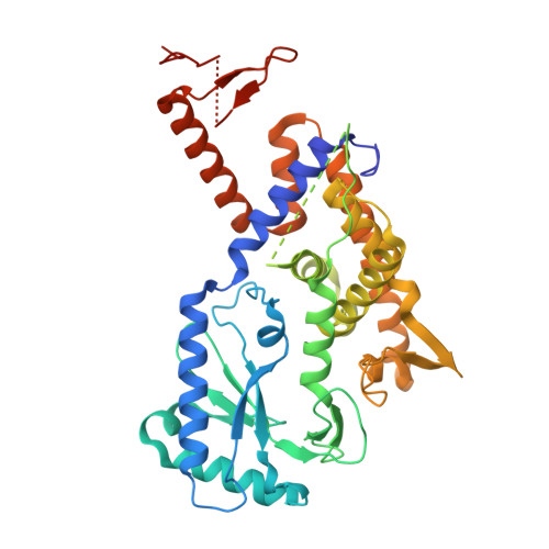
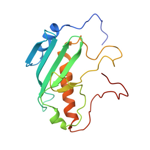
Phage defence system CBASS is regulated by a prokaryotic E2 enzyme that imitates the ubiquitin pathway.

Yan, Y.Xiao, J.Huang, F.Xian, W.Yu, B.Cheng, R.Wu, H.Lu, X.Wang, X.Huang, W.Li, J.Oyejobi, G.K.Robinson, C.V.Wu, H.Wu, D.Liu, X.Wang, L.Zhu, B.

(2024) Nat Microbiol 9: 1566-1578

  • DOI: https://doi.org/10.1038/s41564-024-01684-z
  • Primary Citation of Related Structures:  
    8HSB, 8YJY

  • PubMed Abstract: 

    The cyclic-oligonucleotide-based anti-phage signalling system (CBASS) is a type of innate prokaryotic immune system. Composed of a cyclic GMP-AMP synthase (cGAS) and CBASS-associated proteins, CBASS uses cyclic oligonucleotides to activate antiviral immunity. One major class of CBASS contains a homologue of eukaryotic ubiquitin-conjugating enzymes, which is either an E1-E2 fusion or a single E2. However, the functions of single E2s in CBASS remain elusive. Here, using biochemical, genetic, cryo-electron microscopy and mass spectrometry investigations, we discover that the E2 enzyme from Serratia marcescens regulates cGAS by imitating the ubiquitination cascade. This includes the processing of the cGAS C terminus, conjugation of cGAS to a cysteine residue, ligation of cGAS to a lysine residue, cleavage of the isopeptide bond and poly-cGASylation. The poly-cGASylation activates cGAS to produce cGAMP, which acts as an antiviral signal and leads to cell death. Thus, our findings reveal a unique regulatory role of E2 in CBASS.


  • Organizational Affiliation
    • Key Laboratory of Molecular Biophysics, the Ministry of Education, College of Life Science and Technology, Huazhong University of Science and Technology, Wuhan, China.

Macromolecules
Find similar proteins by:  (by identity cutoff)  |  3D Structure
Entity ID: 1
MoleculeChains Sequence LengthOrganismDetailsImage
SmCdnG407Serratia marcescensMutation(s): 0 
UniProt
Find proteins for A0AAT8XUL1 (Serratia marcescens)
Explore A0AAT8XUL1 
Go to UniProtKB:  A0AAT8XUL1
Entity Groups  
Sequence Clusters30% Identity50% Identity70% Identity90% Identity95% Identity100% Identity
UniProt GroupA0AAT8XUL1
Sequence Annotations
Expand
  • Reference Sequence
Find similar proteins by:  (by identity cutoff)  |  3D Structure
Entity ID: 2
MoleculeChains Sequence LengthOrganismDetailsImage
Type VI secretion protein162Serratia marcescensMutation(s): 0 
Gene Names: AR325_02475AR325_06795
UniProt
Find proteins for A0AAT8XUL2 (Serratia marcescens)
Explore A0AAT8XUL2 
Go to UniProtKB:  A0AAT8XUL2
Entity Groups  
Sequence Clusters30% Identity50% Identity70% Identity90% Identity95% Identity100% Identity
UniProt GroupA0AAT8XUL2
Sequence Annotations
Expand
  • Reference Sequence
Experimental Data & Validation

Experimental Data

  • Method: ELECTRON MICROSCOPY
  • Resolution: 3.30 Å
  • Aggregation State: PARTICLE 
  • Reconstruction Method: SINGLE PARTICLE 

Structure Validation

View Full Validation Report


Entry History & Funding Information

Deposition Data


Funding OrganizationLocationGrant Number
Not funded--

Revision History  (Full details and data files)

  • Version 1.0: 2024-05-01
    Type: Initial release
  • Version 1.1: 2024-06-19
    Changes: Database references