


Structure of the PGK1 from Biortus.
Wang, F., Cheng, W., Lv, Z., Ju, C., Wang, J.To be published.
Experimental Data Snapshot
Starting Model: experimental
View more details
wwPDB Validation 3D Report Full Report

Entity ID: 1 | |||||
|---|---|---|---|---|---|
| Molecule | Chains | Sequence Length | Organism | Details | Image |
| Phosphoglycerate kinase 1 | 416 | Homo sapiens | Mutation(s): 0 Gene Names: PGK1, PGKA, MIG10, OK/SW-cl.110 EC: 2.7.2.3 (PDB Primary Data), 2.7.11.1 (UniProt) | 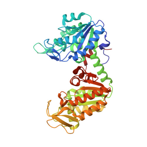 | |
UniProt & NIH Common Fund Data Resources | |||||
PHAROS: P00558 GTEx: ENSG00000102144 | |||||
Entity Groups | |||||
| Sequence Clusters | 30% Identity50% Identity70% Identity90% Identity95% Identity100% Identity | ||||
| UniProt Group | P00558 | ||||
Sequence AnnotationsExpand | |||||
Reference Sequence | |||||
| Ligands 1 Unique | |||||
|---|---|---|---|---|---|
| ID | Chains | Name / Formula / InChI Key | 2D Diagram | 3D Interactions | |
| EDO Download:Ideal Coordinates CCD File | B [auth A], C [auth A] | 1,2-ETHANEDIOL C2 H6 O2 LYCAIKOWRPUZTN-UHFFFAOYSA-N |  | ||
| Length ( Å ) | Angle ( ˚ ) |
|---|---|
| a = 61.591 | α = 90 |
| b = 70.058 | β = 90 |
| c = 95.681 | γ = 90 |
| Software Name | Purpose |
|---|---|
| REFMAC | refinement |
| XDS | data reduction |
| Aimless | data scaling |
| PHASER | phasing |


