8YAD | pdb_00008yad

structure of SPG11-SPG15 complex


Experimental Data Snapshot

  • Method: ELECTRON MICROSCOPY
  • Resolution: 4.02 Å
  • Aggregation State: PARTICLE 
  • Reconstruction Method: SINGLE PARTICLE 

wwPDB Validation   3D Report Full Report


This is version 1.3 of the entry. See complete history


Literature

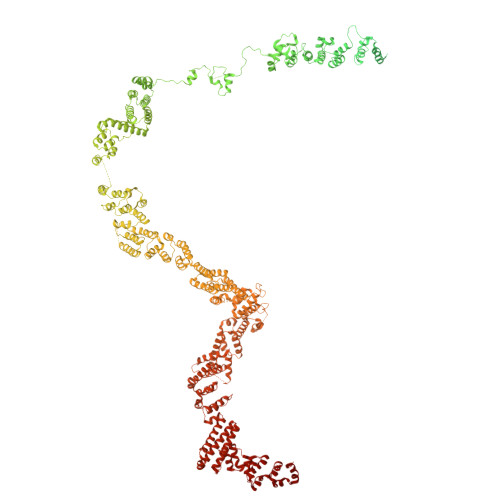
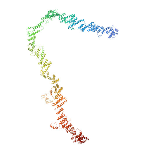
Structural basis for membrane remodeling by the AP5-SPG11-SPG15 complex.

Mai, X.Wang, Y.Wang, X.Liu, M.Teng, F.Liu, Z.Su, M.Y.Stjepanovic, G.

(2025) Nat Struct Mol Biol 32: 1334-1346

  • DOI: https://doi.org/10.1038/s41594-025-01500-0
  • Primary Citation of Related Structures:  
    8YAB, 8YAD, 8YAH

  • PubMed Abstract: 

    The human spastizin (spastic paraplegia 15, SPG15) and spatacsin (spastic paraplegia 11, SPG11) complex is involved in the formation of lysosomes, and mutations in these two proteins are linked with hereditary autosomal-recessive spastic paraplegia. SPG11-SPG15 can cooperate with the fifth adaptor protein complex (AP5) involved in membrane sorting of late endosomes. We employed cryogenic-electron microscopy and in silico predictions to investigate the structural assemblies of the SPG11-SPG15 and AP5-SPG11-SPG15 complexes. The W-shaped SPG11-SPG15 intertwined in a head-to-head fashion, and the N-terminal region of SPG11 is required for AP5 complex interaction and assembly. The AP5 complex is in a super-open conformation. Our findings reveal that the AP5-SPG11-SPG15 complex can bind PI3P molecules, sense membrane curvature and drive membrane remodeling in vitro. These studies provide insights into the structure and function of the spastic paraplegia AP5-SPG11-SPG15 complex, which is essential for the initiation of autolysosome tubulation.


  • Organizational Affiliation
    • Kobilka Institute of Innovative Drug Discovery, School of Medicine, The Chinese University of Hong Kong, Shenzhen, Guangdong, China.

Macromolecules
Find similar proteins by:  (by identity cutoff)  |  3D Structure
Entity ID: 1
MoleculeChains Sequence LengthOrganismDetailsImage
SpatacsinA [auth B]2,443Homo sapiensMutation(s): 0 
Gene Names: SPG11
UniProt & NIH Common Fund Data Resources
Find proteins for Q96JI7 (Homo sapiens)
Explore Q96JI7 
Go to UniProtKB:  Q96JI7
PHAROS:  Q96JI7
GTEx:  ENSG00000104133 
Entity Groups  
Sequence Clusters30% Identity50% Identity70% Identity90% Identity95% Identity100% Identity
UniProt GroupQ96JI7
Sequence Annotations
Expand
  • Reference Sequence
Find similar proteins by:  (by identity cutoff)  |  3D Structure
Entity ID: 2
MoleculeChains Sequence LengthOrganismDetailsImage
Zinc finger FYVE domain-containing protein 26B [auth C]2,539Homo sapiensMutation(s): 0 
Gene Names: ZFYVE26KIAA0321
UniProt & NIH Common Fund Data Resources
Find proteins for Q68DK2 (Homo sapiens)
Explore Q68DK2 
Go to UniProtKB:  Q68DK2
PHAROS:  Q68DK2
GTEx:  ENSG00000072121 
Entity Groups  
Sequence Clusters30% Identity50% Identity70% Identity90% Identity95% Identity100% Identity
UniProt GroupQ68DK2
Sequence Annotations
Expand
  • Reference Sequence
Experimental Data & Validation

Experimental Data

  • Method: ELECTRON MICROSCOPY
  • Resolution: 4.02 Å
  • Aggregation State: PARTICLE 
  • Reconstruction Method: SINGLE PARTICLE 
EM Software:
TaskSoftware PackageVersion
MODEL REFINEMENTPHENIX

Structure Validation

View Full Validation Report


Entry History & Funding Information

Deposition Data

  • Released Date: 2025-03-26 
  • Deposition Author(s): Su, M.-Y.

Funding OrganizationLocationGrant Number
Other governmentChina--

Revision History  (Full details and data files)

  • Version 1.0: 2025-03-26
    Type: Initial release
  • Version 1.1: 2025-04-16
    Changes: Data collection, Database references
  • Version 1.2: 2025-07-02
    Changes: Data collection
  • Version 1.3: 2025-08-27
    Changes: Data collection, Database references