


Crystal structure of CP2c/LSF DNA-binding domain
Park, J., Lee, S.H., Lee, S.H., Kim, S.J., Lee, S.I., Won, H.S., Lee, S.J.To be published.
Experimental Data Snapshot
wwPDB Validation 3D Report Full Report

Entity ID: 1 | |||||
|---|---|---|---|---|---|
| Molecule | Chains | Sequence Length | Organism | Details | Image |
| Alpha-globin transcription factor CP2 | 222 | Homo sapiens | Mutation(s): 0 Gene Names: Tfcp2, Tcfcp2 | 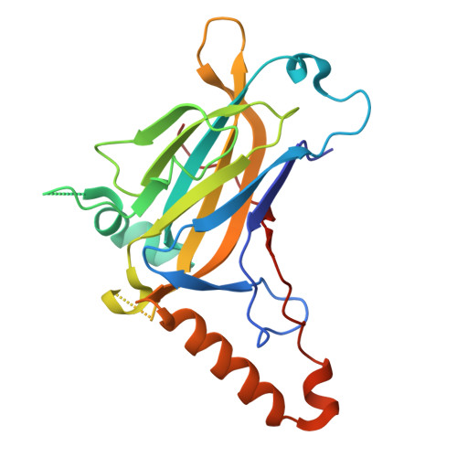 | |
UniProt | |||||
Entity Groups | |||||
| Sequence Clusters | 30% Identity50% Identity70% Identity90% Identity95% Identity100% Identity | ||||
| UniProt Group | Q9ERA0 | ||||
Sequence AnnotationsExpand | |||||
Reference Sequence | |||||
| Length ( Å ) | Angle ( ˚ ) |
|---|---|
| a = 57.093 | α = 90 |
| b = 57.093 | β = 90 |
| c = 130.642 | γ = 90 |
| Software Name | Purpose |
|---|---|
| PHENIX | refinement |
| Coot | model building |
| XDS | data scaling |
| PHENIX | phasing |
| Funding Organization | Location | Grant Number |
|---|---|---|
| National Research Foundation (NRF, Korea) | Korea, Republic Of | -- |


