8Y6I | pdb_00008y6i

P-glycoprotein in complex with UIC2 Fab and triple elacridar molecules in nanodisc


Experimental Data Snapshot

  • Method: ELECTRON MICROSCOPY
  • Resolution: 2.54 Å
  • Aggregation State: PARTICLE 
  • Reconstruction Method: SINGLE PARTICLE 

Starting Model: experimental
View more details

wwPDB Validation 3D Report Full Report

Validation slider image for 8Y6I

This is version 1.2 of the entry. See complete history

Literature

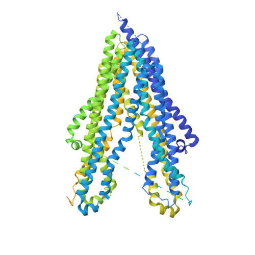
Cryo-EM structure of P-glycoprotein bound to triple elacridar inhibitor molecules.

Hamaguchi-Suzuki, N.Adachi, N.Moriya, T.Yasuda, S.Kawasaki, M.Suzuki, K.Ogasawara, S.Anzai, N.Senda, T.Murata, T.

(2024) Biochem Biophys Res Commun 709: 149855-149855

  • DOI: https://doi.org/10.1016/j.bbrc.2024.149855
  • Primary Citation Related Structures: 
    8Y6H, 8Y6I

  • PubMed Abstract: 

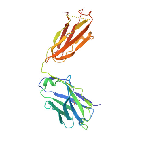
    P-glycoprotein (P-gp) is an ATP-binding cassette transporter known for its roles in expelling xenobiotic compounds from cells and contributing to cellular drug resistance through multidrug efflux. This mechanism is particularly problematic in cancer cells, where it diminishes the therapeutic efficacy of anticancer drugs. P-gp inhibitors, such as elacridar, have been developed to circumvent the decrease in drug efficacy due to P-gp efflux. An earlier study reported the cryo-EM structure of human P-gp-Fab (MRK-16) complex bound by two elacridar molecules, at a resolution of 3.6 Å. In this study, we have obtained a higher resolution (2.5 Å) structure of the P-gp- Fab (UIC2) complex bound by three elacridar molecules. This finding, which exposes a larger space for compound-binding sites than previously acknowledged, has significant implications for the development of more selective inhibitors and enhances our understanding of the compound recognition mechanism of P-gp.


  • Organizational Affiliation
    • Department of Pharmacology, Chiba University Graduate School of Medicine, 1-8-1 Inohana, Chuo, Chiba, 260-8670, Japan; Department of Chemistry, Graduate School of Science, Chiba University, 1-33 Yayoi-cho, Inage, Chiba, 263-8522, Japan.

Macromolecule Content 

  • Total Structure Weight: 226.32 kDa 
  • Atom Count: 8,406 
  • Modeled Residue Count: 1,041 
  • Deposited Residue Count: 1,979 
  • Unique protein chains: 3

Macromolecules

Find similar proteins by:|  3D Structure
Entity ID: 1
MoleculeChains  Sequence LengthOrganismDetailsImage
ATP-dependent translocase ABCB1,mNeonGreen1,534Homo sapiensMutation(s): 0 
Gene Names: ABCB1MDR1PGY1blFP-Y3
EC: 7.6.2.2 (PDB Primary Data), 7.6.2.1 (PDB Primary Data)
Membrane Entity: Yes 
UniProt & NIH Common Fund Data Resources
Find proteins for P08183 (Homo sapiens)
Explore P08183 
Go to UniProtKB:  P08183
PHAROS:  P08183
GTEx:  ENSG00000085563 
Entity Groups
Sequence Clusters30% Identity50% Identity70% Identity90% Identity95% Identity100% Identity
UniProt GroupP08183
Sequence Annotations
Expand
Reference Sequence
Find similar proteins by:|  3D Structure
Entity ID: 2
MoleculeChains  Sequence LengthOrganismDetailsImage
UIC2 Fab light chain220Mus musculusMutation(s): 0 
Find similar proteins by:|  3D Structure
Entity ID: 3
MoleculeChains  Sequence LengthOrganismDetailsImage
UIC2 Fab heavy chain225Mus musculusMutation(s): 0 

Small Molecules

Ligands 3 Unique
IDChains Name / Formula / InChI Key2D Diagram3D Interactions
3PE

Query on 3PE


Download:Ideal Coordinates CCD File
O [auth A]1,2-Distearoyl-sn-glycerophosphoethanolamine
C41 H82 N O8 P
LVNGJLRDBYCPGB-LDLOPFEMSA-N
R0Z
(Subject of Investigation/LOI)

Query on R0Z


Download:Ideal Coordinates CCD File
Q [auth A],
R [auth A],
S [auth A]
elacridar
C34 H33 N3 O5
OSFCMRGOZNQUSW-UHFFFAOYSA-N
CLR

Query on CLR


Download:Ideal Coordinates CCD File
D [auth A]
E [auth A]
F [auth A]
G [auth A]
H [auth A]
D [auth A],
E [auth A],
F [auth A],
G [auth A],
H [auth A],
I [auth A],
J [auth A],
K [auth A],
L [auth A],
M [auth A],
N [auth A],
P [auth A]
CHOLESTEROL
C27 H46 O
HVYWMOMLDIMFJA-DPAQBDIFSA-N
Binding Affinity Annotations 
IDSourceBinding Affinity
R0Z BindingDB:  8Y6I Ki: min: 40, max: 440 (nM) from 2 assay(s)
IC50: min: 4.9, max: 2000 (nM) from 4 assay(s)
EC50: min: 14, max: 2000 (nM) from 2 assay(s)

Experimental Data & Validation

Experimental Data

  • Method: ELECTRON MICROSCOPY
  • Resolution: 2.54 Å
  • Aggregation State: PARTICLE 
  • Reconstruction Method: SINGLE PARTICLE 
EM Software:
TaskSoftware PackageVersion
RECONSTRUCTIONcryoSPARC4.2.1
MODEL REFINEMENTPHENIX1.20.1

Structure Validation

View Full Validation Report


Entry History 

& Funding Information

Deposition Data


Funding OrganizationLocationGrant Number
Japan Agency for Medical Research and Development (AMED)JapanJP21am0101083
Japan Agency for Medical Research and Development (AMED)JapanJP23ama121013
Japan Society for the Promotion of Science (JSPS)Japan18H05425

Revision History  (Full details and data files)

  • Version 1.0: 2024-04-17
    Type: Initial release
  • Version 1.1: 2024-11-13
    Changes: Data collection, Structure summary
  • Version 1.2: 2024-11-27
    Changes: Data collection