8Y56 | pdb_00008y56

Cryo-EM reveals cholesterol binding in the lysosomal GPCR-like protein LYCHOS


Experimental Data Snapshot

  • Method: ELECTRON MICROSCOPY
  • Resolution: 2.83 Å
  • Aggregation State: PARTICLE 
  • Reconstruction Method: SINGLE PARTICLE 

wwPDB Validation 3D Report Full Report

Validation slider image for 8Y56

This is version 1.2 of the entry. See complete history. 

Literature

Cryo-EM reveals cholesterol binding in the lysosomal GPCR-like protein LYCHOS.

Zhao, J., Shen, Q., Yong, X., Li, X., Tian, X., Sun, S., Xu, Z., Zhang, X., Zhang, L., Yang, H., Shao, Z., Xu, H., Jiang, Y., Zhang, Y., Yan, W.

(2025) Nat Struct Mol Biol 32: 896-904

  • DOI: https://doi.org/10.1038/s41594-024-01470-9
  • Primary Citation Related Structures: 
    8Y56

  • PubMed Abstract: 

    Cholesterol plays a pivotal role in modulating the activity of mechanistic target of rapamycin complex 1 (mTOR1), thereby regulating cell growth and metabolic homeostasis. LYCHOS, a lysosome-localized G-protein-coupled receptor-like protein, emerges as a cholesterol sensor and is capable of transducing the cholesterol signal to affect the mTORC1 function. However, the precise mechanism by which LYCHOS recognizes cholesterol remains unknown. Here, using cryo-electron microscopy, we determined the three-dimensional structural architecture of LYCHOS in complex with cholesterol molecules, revealing a unique arrangement of two sequential structural domains. Through a comprehensive analysis of this structure, we elucidated the specific structural features of these two domains and their collaborative role in the process of cholesterol recognition by LYCHOS.


  • Organizational Affiliation: 
    • Division of Nephrology and Kidney Research Institute, State Key Laboratory of Biotherapy, West China Hospital, Sichuan University, Chengdu, China.

Macromolecule Content 

  • Total Structure Weight: 96.55 kDa 
  • Atom Count: 4,796 
  • Modeled Residue Count: 586 
  • Deposited Residue Count: 840 
  • Unique protein chains: 1

Macromolecules

Find similar proteins by:|  3D Structure
Entity ID: 1
MoleculeChains  Sequence LengthOrganismDetailsImage
Lysosomal cholesterol signaling proteinA [auth R]840Homo sapiensMutation(s): 0 
Gene Names: GPR155, PGR22
Membrane Entity: Yes 
UniProt & NIH Common Fund Data Resources
Find proteins for Q7Z3F1 (Homo sapiens)
Explore Q7Z3F1 
Go to UniProtKB:  Q7Z3F1
PHAROS:  Q7Z3F1
GTEx:  ENSG00000163328 
Entity Groups
Sequence Clusters30% Identity50% Identity70% Identity90% Identity95% Identity100% Identity
UniProt GroupQ7Z3F1
Sequence Annotations
Expand
Reference Sequence

Small Molecules

Ligands 3 Unique
IDChains Name / Formula / InChI Key2D Diagram3D Interactions
DKB

Query on DKB


Download:Ideal Coordinates CCD File
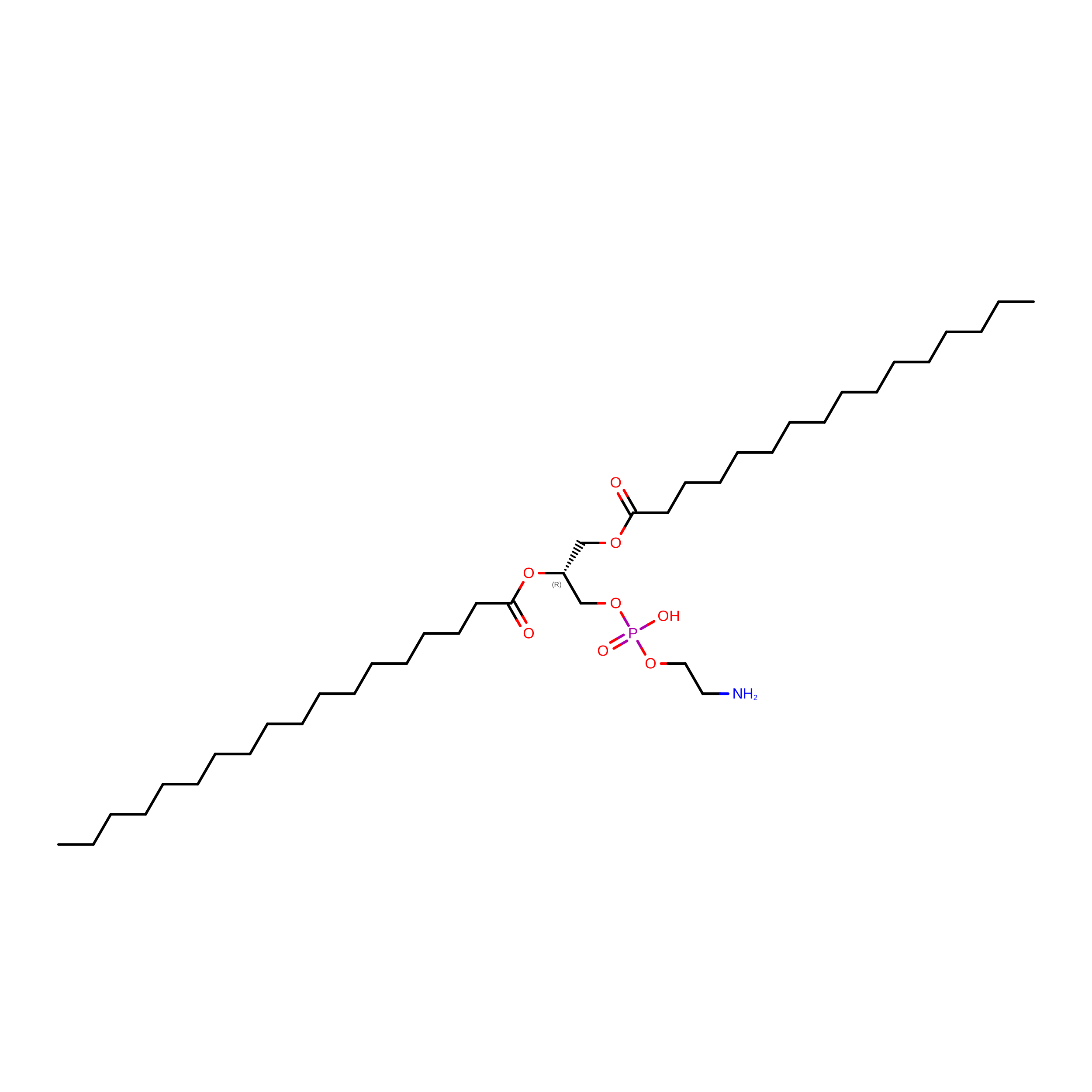
H [auth R][(2R)-1-[2-azanylethoxy(oxidanyl)phosphoryl]oxy-3-hexadecanoyloxy-propan-2-yl] octadecanoate
C39 H78 N O8 P
RPJZYOHZALDGKI-DIPNUNPCSA-N
CLR
(Subject of Investigation/LOI)

Query on CLR


Download:Ideal Coordinates CCD File
C [auth R],
D [auth R],
E [auth R],
F [auth R],
G [auth R]
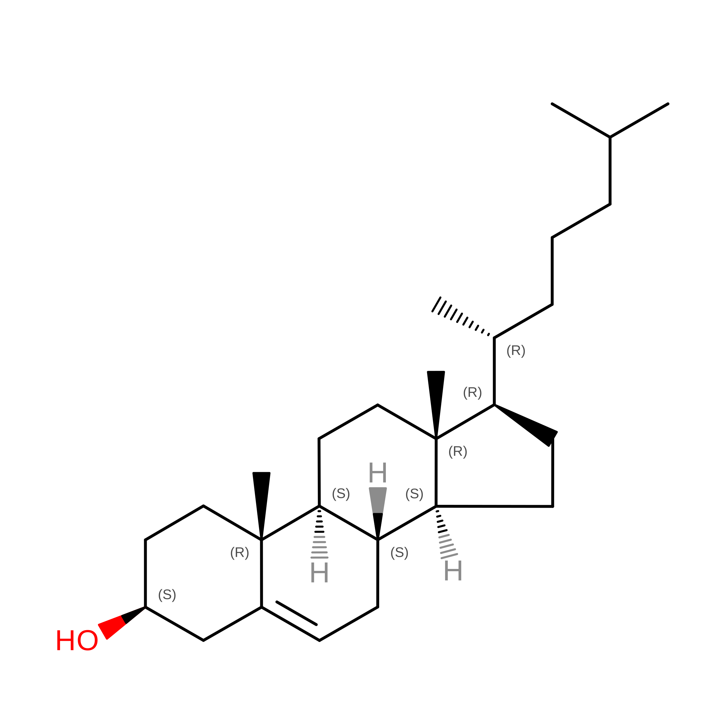
CHOLESTEROL
C27 H46 O
HVYWMOMLDIMFJA-DPAQBDIFSA-N
NA

Query on NA


Download:Ideal Coordinates CCD File
B [auth R]SODIUM ION
Na
FKNQFGJONOIPTF-UHFFFAOYSA-N

Experimental Data & Validation

Experimental Data

  • Method: ELECTRON MICROSCOPY
  • Resolution: 2.83 Å
  • Aggregation State: PARTICLE 
  • Reconstruction Method: SINGLE PARTICLE 
EM Software:
TaskSoftware PackageVersion
MODEL REFINEMENTPHENIX

Structure Validation

View Full Validation Report


Entry History 

& Funding Information

Deposition Data


Funding OrganizationLocationGrant Number
Not funded--

Revision History  (Full details and data files)

  • Version 1.0: 2025-01-29
    Type: Initial release
  • Version 1.1: 2025-06-04
    Changes: Data collection, Database references
  • Version 1.2: 2025-07-23
    Changes: Data collection