 Download Mendeley
Crystal structure of ferric iron-binding protein (FecB) from Vibrio alginolyticus in complex with ferric citrate
Jiang, J.Y., Lu, P., Nagata, K.To be published.
Experimental Data Snapshot
Starting Model: in silico
View more details
wwPDB Validation 3D Report Full Report

Entity ID: 1 | |||||
|---|---|---|---|---|---|
| Molecule | Chains | Sequence Length | Organism | Details | Image |
| ABC transporter substrate-binding protein | 271 | Vibrio alginolyticus | Mutation(s): 0 Gene Names: HKB16_09180 |  | |
UniProt | |||||
Find proteins for A0A7Y0SGI3 (Vibrio parahaemolyticus) Explore A0A7Y0SGI3 Go to UniProtKB: A0A7Y0SGI3 | |||||
Entity Groups | |||||
| Sequence Clusters | 30% Identity50% Identity70% Identity90% Identity95% Identity100% Identity | ||||
| UniProt Group | A0A7Y0SGI3 | ||||
Sequence AnnotationsExpand | |||||
Reference Sequence | |||||
| Ligands 4 Unique | |||||
|---|---|---|---|---|---|
| ID | Chains | Name / Formula / InChI Key | 2D Diagram | 3D Interactions | |
| FLC Download:Ideal Coordinates CCD File | F [auth A], G [auth A] | CITRATE ANION C6 H5 O7 KRKNYBCHXYNGOX-UHFFFAOYSA-K | 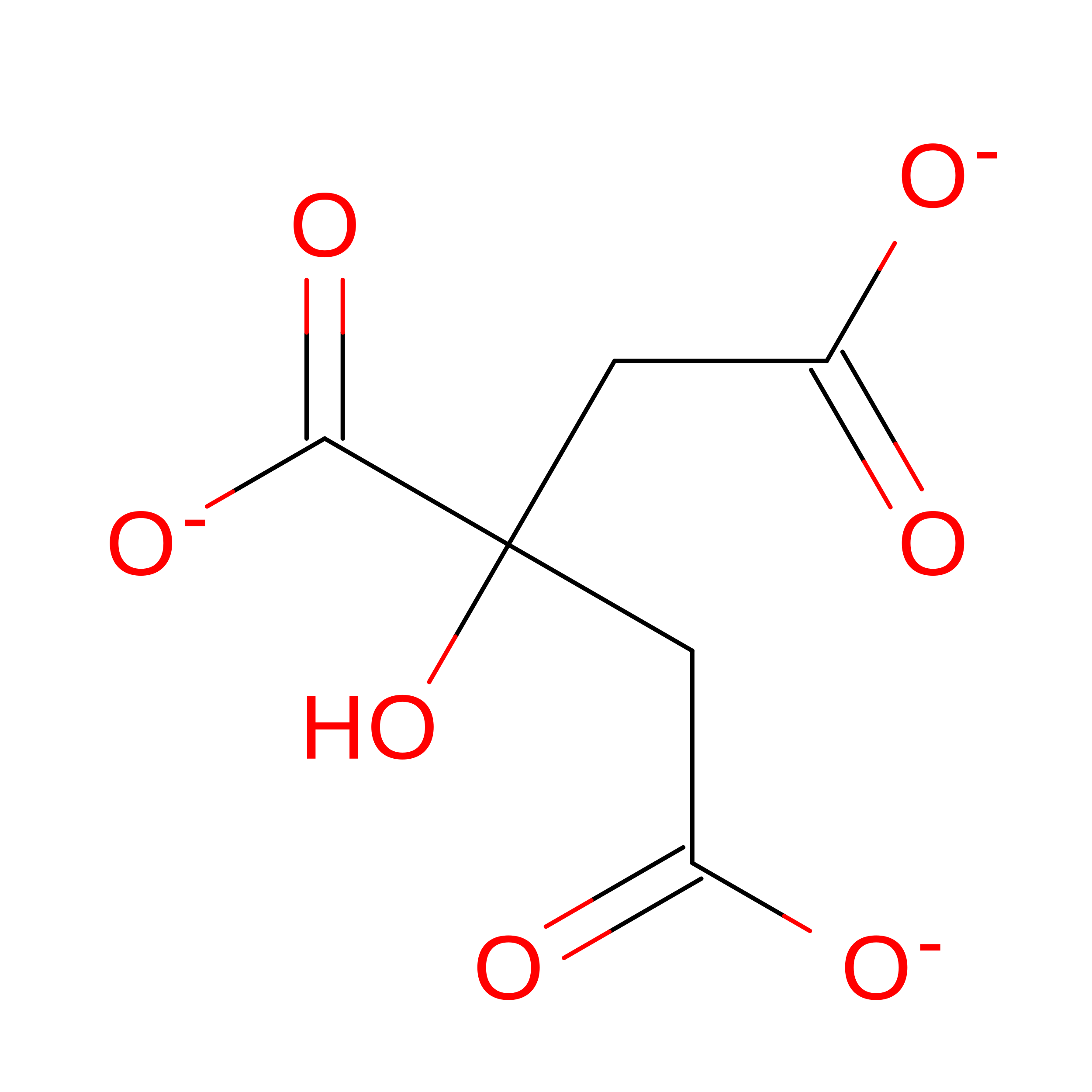 | ||
| PEG Download:Ideal Coordinates CCD File | E [auth A] | DI(HYDROXYETHYL)ETHER C4 H10 O3 MTHSVFCYNBDYFN-UHFFFAOYSA-N |  | ||
| GOL Download:Ideal Coordinates CCD File | B [auth A], C [auth A], D [auth A] | GLYCEROL C3 H8 O3 PEDCQBHIVMGVHV-UHFFFAOYSA-N |  | ||
| FE Download:Ideal Coordinates CCD File | H [auth A] | FE (III) ION Fe VTLYFUHAOXGGBS-UHFFFAOYSA-N | 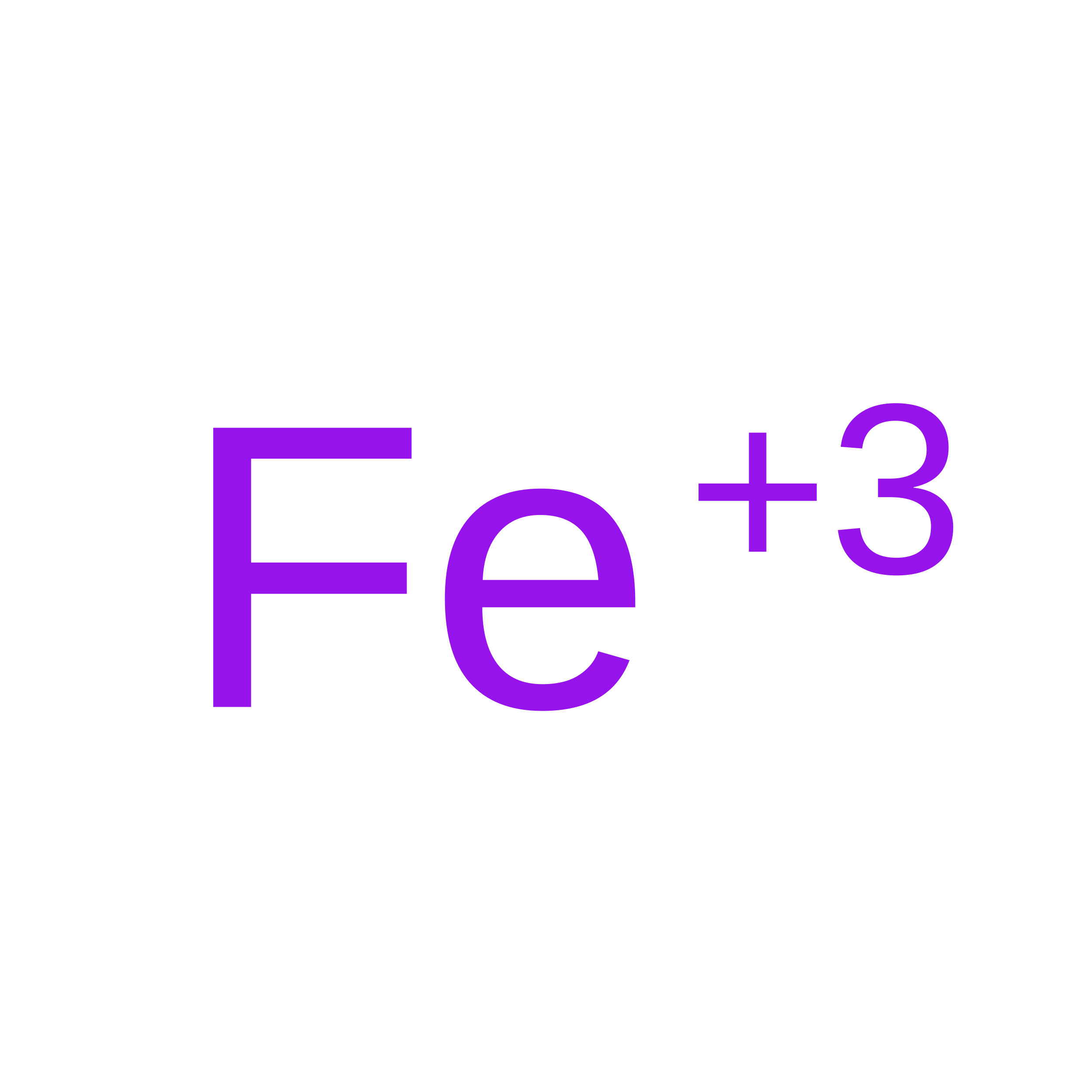 | ||
| Length ( Å ) | Angle ( ˚ ) |
|---|---|
| a = 100.497 | α = 90 |
| b = 100.497 | β = 90 |
| c = 205.983 | γ = 90 |
| Software Name | Purpose |
|---|---|
| PHENIX | refinement |
| HKL-2000 | data reduction |
| REFMAC | phasing |
| HKL-2000 | data collection |
| XDS | data scaling |
| Funding Organization | Location | Grant Number |
|---|---|---|
| Japan Society for the Promotion of Science (JSPS) | Japan | 19H05771 |
| Japan Society for the Promotion of Science (JSPS) | Japan | JP20K22561 |
| Japan Science and Technology | Japan | JPMJSP2108 |
| Other private | Japan Science Society 2023-4016 |
