8XGM | pdb_00008xgm

Cryo-EM structure of human GPR1 bound to chemerin


Experimental Data Snapshot

  • Method: ELECTRON MICROSCOPY
  • Resolution: 3.29 Å
  • Aggregation State: PARTICLE 
  • Reconstruction Method: SINGLE PARTICLE 

wwPDB Validation   3D Report Full Report


This is version 1.3 of the entry. See complete history


Literature

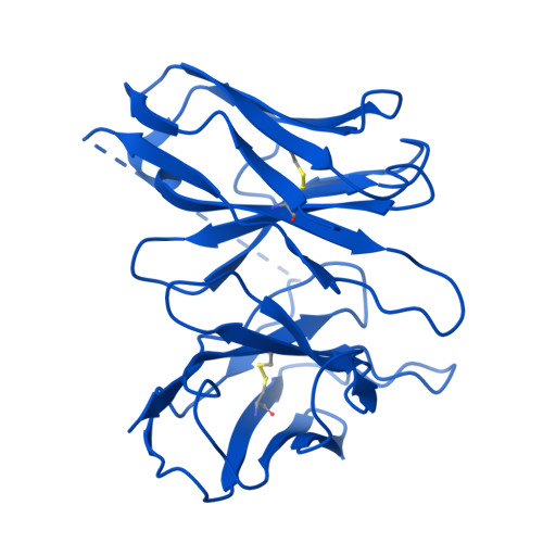
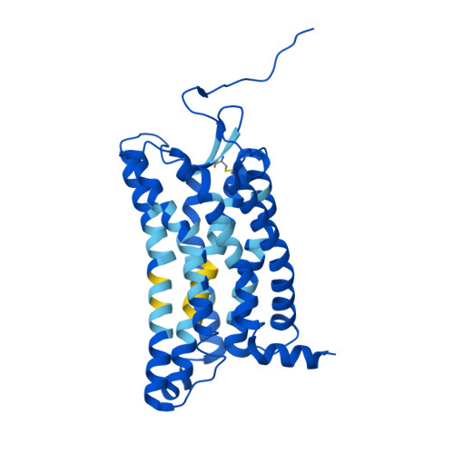
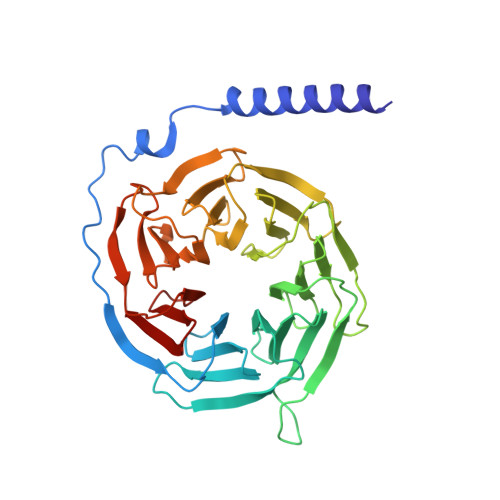
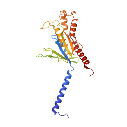
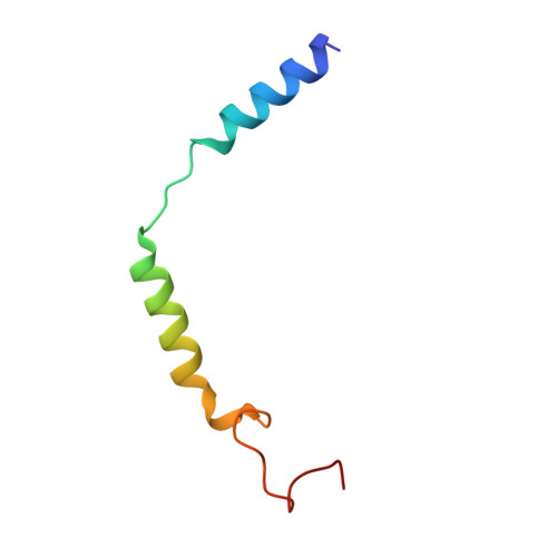
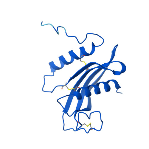
Structure of G protein-coupled receptor GPR1 bound to full-length chemerin adipokine reveals a chemokine-like reverse binding mode.

Liu, A.Liu, Y.Chen, G.Lyu, W.Ye, F.Wang, J.Liao, Q.Zhu, L.Du, Y.Ye, R.D.

(2024) PLoS Biol 22: e3002838-e3002838

  • DOI: https://doi.org/10.1371/journal.pbio.3002838
  • Primary Citation of Related Structures:  
    8JJP, 8XGM

  • PubMed Abstract: 

    Chemerin is an adipokine with chemotactic activity to a subset of leukocytes. Chemerin binds to 3 G protein-coupled receptors, including chemokine-like receptor 1 (CMKLR1), G protein-coupled receptor 1 (GPR1), and C-C chemokine receptor-like 2 (CCRL2). Here, we report that GPR1 is capable of Gi signaling when stimulated with full-length chemerin or its C-terminal nonapeptide (C9, YFPGQFAFS). We present high-resolution cryo-EM structures of Gi-coupled GPR1 bound to full-length chemerin and to the C9 peptide, respectively. C9 insertion into the transmembrane (TM) binding pocket is both necessary and sufficient for GPR1 signaling, whereas the full-length chemerin uses its bulky N-terminal core for interaction with a β-strand located at the N-terminus of GPR1. This interaction involves multiple β-strands of full-length chemerin, forming a β-sheet that serves as a "lid" for the TM binding pocket and is energetically expensive to remove as indicated by molecular dynamics simulations with free energy landscape analysis. Combining results from functional assays, our structural model explains why C9 is an activating peptide at GPR1 and how the full-length chemerin uses a "two-site" model for enhanced interaction with GPR1.


  • Organizational Affiliation
    • Kobilka Institute of Innovative Drug Discovery, School of Medicine, The Chinese University of Hong Kong, Shenzhen, China.

Macromolecules
Find similar proteins by:  (by identity cutoff)  |  3D Structure
Entity ID: 1
MoleculeChains Sequence LengthOrganismDetailsImage
scFV16A [auth S]247Vicugna pacosMutation(s): 0 
Entity Groups  
Sequence Clusters30% Identity50% Identity70% Identity90% Identity95% Identity100% Identity
Sequence Annotations
Expand
  • Reference Sequence
Find similar proteins by:  (by identity cutoff)  |  3D Structure
Entity ID: 2
MoleculeChains Sequence LengthOrganismDetailsImage
G-protein coupled receptor 1B [auth A]306Homo sapiensMutation(s): 0 
Gene Names: GPR1
Membrane Entity: Yes 
UniProt & NIH Common Fund Data Resources
Find proteins for P46091 (Homo sapiens)
Explore P46091 
Go to UniProtKB:  P46091
PHAROS:  P46091
GTEx:  ENSG00000183671 
Entity Groups  
Sequence Clusters30% Identity50% Identity70% Identity90% Identity95% Identity100% Identity
UniProt GroupP46091
Sequence Annotations
Expand
  • Reference Sequence
Find similar proteins by:  (by identity cutoff)  |  3D Structure
Entity ID: 3
MoleculeChains Sequence LengthOrganismDetailsImage
Guanine nucleotide-binding protein G(I)/G(S)/G(T) subunit beta-1C [auth B]340Homo sapiensMutation(s): 0 
Gene Names: GNB1
UniProt & NIH Common Fund Data Resources
Find proteins for P62873 (Homo sapiens)
Explore P62873 
Go to UniProtKB:  P62873
PHAROS:  P62873
GTEx:  ENSG00000078369 
Entity Groups  
Sequence Clusters30% Identity50% Identity70% Identity90% Identity95% Identity100% Identity
UniProt GroupP62873
Sequence Annotations
Expand
  • Reference Sequence
Find similar proteins by:  (by identity cutoff)  |  3D Structure
Entity ID: 4
MoleculeChains Sequence LengthOrganismDetailsImage
Guanine nucleotide-binding protein G(i) subunit alpha-1D [auth C]354Homo sapiensMutation(s): 2 
Gene Names: GNAI1
EC: 3.6.5
Membrane Entity: Yes 
UniProt & NIH Common Fund Data Resources
Find proteins for P63096 (Homo sapiens)
Explore P63096 
Go to UniProtKB:  P63096
PHAROS:  P63096
GTEx:  ENSG00000127955 
Entity Groups  
Sequence Clusters30% Identity50% Identity70% Identity90% Identity95% Identity100% Identity
UniProt GroupP63096
Sequence Annotations
Expand
  • Reference Sequence
Find similar proteins by:  (by identity cutoff)  |  3D Structure
Entity ID: 5
MoleculeChains Sequence LengthOrganismDetailsImage
Guanine nucleotide-binding protein subunit gammaE [auth G]55Homo sapiensMutation(s): 0 
Gene Names: GNG2
Membrane Entity: Yes 
UniProt & NIH Common Fund Data Resources
Find proteins for P59768 (Homo sapiens)
Explore P59768 
Go to UniProtKB:  P59768
PHAROS:  P59768
GTEx:  ENSG00000186469 
Entity Groups  
Sequence Clusters30% Identity50% Identity70% Identity90% Identity95% Identity100% Identity
UniProt GroupP59768
Sequence Annotations
Expand
  • Reference Sequence
Find similar proteins by:  (by identity cutoff)  |  3D Structure
Entity ID: 6
MoleculeChains Sequence LengthOrganismDetailsImage
Retinoic acid receptor responder protein 2F [auth D]137Homo sapiensMutation(s): 0 
Gene Names: RARRES2
Membrane Entity: Yes 
UniProt & NIH Common Fund Data Resources
Find proteins for Q99969 (Homo sapiens)
Explore Q99969 
Go to UniProtKB:  Q99969
PHAROS:  Q99969
GTEx:  ENSG00000106538 
Entity Groups  
Sequence Clusters30% Identity50% Identity70% Identity90% Identity95% Identity100% Identity
UniProt GroupQ99969
Sequence Annotations
Expand
  • Reference Sequence
Small Molecules
Ligands 1 Unique
IDChains Name / Formula / InChI Key2D Diagram3D Interactions
CLR
Query on CLR

Download Ideal Coordinates CCD File 
G [auth A]CHOLESTEROL
C27 H46 O
HVYWMOMLDIMFJA-DPAQBDIFSA-N
Experimental Data & Validation

Experimental Data

  • Method: ELECTRON MICROSCOPY
  • Resolution: 3.29 Å
  • Aggregation State: PARTICLE 
  • Reconstruction Method: SINGLE PARTICLE 
EM Software:
TaskSoftware PackageVersion
MODEL REFINEMENTPHENIX

Structure Validation

View Full Validation Report


Entry History & Funding Information

Deposition Data


Funding OrganizationLocationGrant Number
Not funded--

Revision History  (Full details and data files)

  • Version 1.0: 2024-08-21
    Type: Initial release
  • Version 1.1: 2024-10-09
    Changes: Data collection, Structure summary
  • Version 1.2: 2025-03-05
    Changes: Data collection, Database references
  • Version 1.3: 2025-07-02
    Changes: Data collection