Complete xylan utilization pathway and regulation mechanisms involved in marine algae degradation by cosmopolitan marine and human gut microbiota.
Sun, H.N., Chen, X.L., Wang, Y., Zhu, Y.P., Teng, Z.J., Cao, H.Y., Xu, T.T., Chen, Y., Zhang, Y.Z., Zhao, F.(2025) ISME J 19
- PubMed: 40401997 Search on PubMedSearch on PubMed Central
- DOI: https://doi.org/10.1093/ismejo/wraf085
- Primary Citation Related Structures:
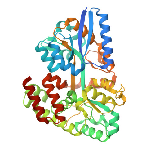
8XBA, 8XBB, 8XBC - PubMed Abstract:
β-1,3-xylan, typically found in marine algae as a major cell wall polysaccharide, represents an overlooked pool of organic carbon in global oceans. Whilst our understanding of microbial catabolism of xylans has improved significantly, particularly from biotransformations of terrestrial plant biomass that are typically composed of β-1,4-xylans, knowledge on how microbes utilize β-1,3-xylan remains limited. Here, we describe the discovery of a complete pathway for β-1,3-xylan catabolism and its regulation in a marine bacterium, Vibrio sp. EA2. The pathway starts with the extracellular decomposition of β-1,3-xylan by two β-1,3-xylanases into β-1,3-xylooligomers, which are mainly internalized by an ATP-binding cassette transporter. The substrate binding protein of this transporter has an L-shaped substrate binding pocket to preferentially bind β-1,3-xylooligomers. Subsequently, two intracellular β-1,3-xylosidases degrade β-1,3-xylooligomers into fermentable xylose. The pathway is activated by a unique regulator with xylose being the effector. This β-1,3-xylan catabolic pathway differs from that of β-1,4-xylan catabolism in enzymes, transporters, and regulators. Bioinformatic analysis suggests that the β-1,3-xylan catabolism pathway is not only prevalent in diverse marine bacteria and cosmopolitan human gut microbiota, such as Bacteroides, but also likely transferred horizontally from algae-degrading marine bacteria to the human gut.
- State Key Laboratory of Microbial Technology, Shandong University, Qingdao 266237, Shandong Province, China.
Organizational Affiliation: