Hemagglutinin double-mutation enhances binding of human-infecting avian influenza virus clade 2.3.4.4b H5Ny to human and SLe X receptors.
Jin, X., Han, P., Wang, Y., Wang, H., Zhang, C., Di Maio, A., Yu, J., Hao, T., Gu, Y., Zhang, Z., Zhang, W., Qi, J., Bi, Y., Zhang, X., Sun, L., Wang, N., Liu, Y., Song, H., Gao, G.F.(2026) EMBO Rep
- PubMed: 42303811 Search on PubMedSearch on PubMed Central
- DOI: https://doi.org/10.1038/s44319-026-00816-2
- Primary Citation Related Structures:
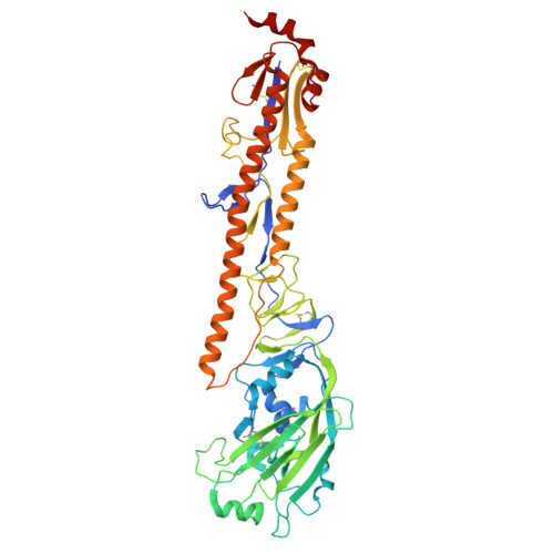
8X26, 8X27, 8X28, 8X29, 8X2C, 8X2D, 8X2F - PubMed Abstract:
Clade 2.3.4.4b H5Ny highly pathogenic avian influenza viruses (HPAIVs) continue to circulate worldwide, posing zoonotic threats, especially with recent cattle outbreaks. The mechanisms by which these viruses adapt to mammalian hosts while maintaining a broad avian tropism remain poorly understood. Here, we demonstrate that two naturally occurring mutations (K222Q and S227R) in the hemagglutinin (HA) of a human-infecting H5N8 strain, first identified in 2020, enhance binding affinity for both α2-6-linked and Sialyl Lewis X (SLe X ) glycans, which may underlie the broad tissue binding and cross-species potential. Structural analyses reveal that these mutations expand receptor specificity for these glycans, which are abundant in the human respiratory tract and duck trachea, providing a possible molecular basis for cross-species transmission. Our findings suggest that clade 2.3.4.4b H5Ny viruses evolved dual receptor specificity as early as the 2020 Russian H5N8 strain, potentially contributing to sporadic human infections and widespread dissemination among birds and mammals.
- CAS Key Laboratory of Pathogen Microbiology and Immunology, Institute of Microbiology, Chinese Academy of Sciences, Beijing, China.
Organizational Affiliation: