8WZC | pdb_00008wzc

NYN domain of human KHNYN complex with RNA


Experimental Data Snapshot

  • Method: X-RAY DIFFRACTION
  • Resolution: 1.93 Å
  • R-Value Free: 
    0.209 (Depositor), 0.220 (DCC) 
  • R-Value Work: 
    0.180 (Depositor), 0.192 (DCC) 

wwPDB Validation 3D Report Full Report

Validation slider image for 8WZC

Ligand Structure Quality Assessment 


This is version 1.0 of the entry. See complete history

Literature

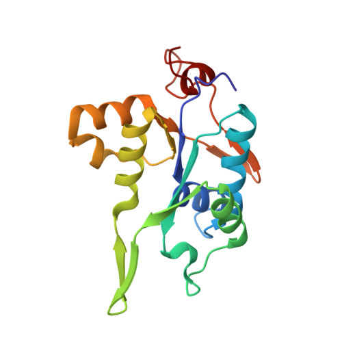
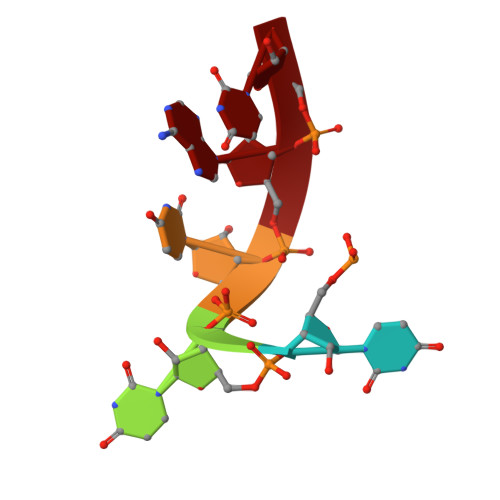
Crystal structure of NYN domain of Human KHNYN in complex with single strand RNA.

Hong, S.Choe, J.

(2024) Biochem Biophys Res Commun 738: 150545-150545

  • DOI: https://doi.org/10.1016/j.bbrc.2024.150545
  • Primary Citation Related Structures: 
    8WZC

  • PubMed Abstract: 

    KHNYN protein with a KH-like domain and a NYN endoribonuclease domain interacts with Zinc-finger antiviral protein (ZAP). ZAP isoforms recognize viral or cellular RNAs and recruit KHNYN to form the ZAP: KHNYN complex. Although the structures of several PIN/NYN domains have been determined, the precise substrate RNA binding mode remains poorly understood. This study presents the crystal structure of a complex of the NYN domain of KHNYN and a 7mer RNA from interferon lambda3 (IFNL3). Our structural analysis reveals that NYN domain of human KHNYN shares structural similarities with other NYN domains of ZC3H12àC proteins. The RNA is bound in the central groove region of the protein, facilitated by interactions including coordination by two Mg 2+ ions, hydrophobic interactions, and hydrogen bonds. In the observed RNA-protein complex, the U 5 , A 6 , and U 7 bases are stacked on top of one another, while U 3 and U 4 bases adopt an "open" conformation (as opposed to base-stacked), forming a U-shaped overall structure. Mutagenesis studies underscore the significance of residues involved in RNA binding for RNase activity. Interestingly, NYN domain of human KHNYN forms a head-to-tail dimer in the crystal, a structural feature also observed in other homologous PIN/NYN proteins, with a residue from the symmetry mate contributing to hydrophobic interactions with the bound RNA.


  • Organizational Affiliation
    • Department of Life Science, University of Seoul, Seoul, 02504, Republic of Korea.

Macromolecule Content 

  • Total Structure Weight: 20.56 kDa 
  • Atom Count: 1,609 
  • Modeled Residue Count: 168 
  • Deposited Residue Count: 168 
  • Unique protein chains: 1
  • Unique nucleic acid chains: 1

Macromolecules


Find similar proteins by:|  3D Structure
Entity ID: 1
MoleculeChains  Sequence LengthOrganismDetailsImage
Protein KHNYN163Homo sapiensMutation(s): 0 
Gene Names: KHNYN
UniProt & NIH Common Fund Data Resources
Find proteins for O15037 (Homo sapiens)
Explore O15037 
Go to UniProtKB:  O15037
PHAROS:  O15037
GTEx:  ENSG00000100441 
Entity Groups
Sequence Clusters30% Identity50% Identity70% Identity90% Identity95% Identity100% Identity
UniProt GroupO15037
Sequence Annotations
Expand
Reference Sequence
Find similar nucleic acids by:  Sequence
Entity ID: 2
MoleculeChains LengthOrganismImage
RNA (5'-R(P*UP*UP*UP*AP*U)-3')B [auth D]5Homo sapiens
Sequence Annotations
Expand
Reference Sequence

Experimental Data & Validation

Experimental Data

  • Method: X-RAY DIFFRACTION
  • Resolution: 1.93 Å
  • R-Value Free:  0.209 (Depositor), 0.220 (DCC) 
  • R-Value Work:  0.180 (Depositor), 0.192 (DCC) 
Space Group: P 41 3 2
Unit Cell:
Length ( Å )Angle ( ˚ )
a = 111.197α = 90
b = 111.197β = 90
c = 111.197γ = 90
Software Package:
Software NamePurpose
REFMACrefinement
HKL-2000data reduction
HKL-2000data scaling
SHELXDphasing

Structure Validation

View Full Validation Report


Ligand Structure Quality Assessment 


Entry History 

& Funding Information

Deposition Data


Funding OrganizationLocationGrant Number
National Research Foundation (NRF, Korea)Korea, Republic Of--

Revision History  (Full details and data files)

  • Version 1.0: 2024-09-11
    Type: Initial release