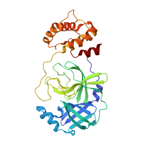
8WSJ | pdb_00008wsj

Crystal structure of SARS-Cov-2 main protease, pH=6.5


Experimental Data Snapshot

  • Method: X-RAY DIFFRACTION
  • Resolution: 1.74 Å
  • R-Value Free: 
    0.233 (Depositor), 0.234 (DCC) 
  • R-Value Work: 
    0.209 (Depositor), 0.209 (DCC) 
  • R-Value Observed: 
    0.210 (Depositor) 

Starting Model: experimental
View more details

wwPDB Validation 3D Report Full Report

Validation slider image for 8WSJ

This is version 1.1 of the entry. See complete history. 

Literature

Crystal structures of SARS-CoV-2 main protease and its complex with PBI-0451.

Jiang, H., Li, W., Zhang, J., Li, J.

(2026) Acta Crystallogr D Struct Biol 82: 1260-1273

  • DOI: https://doi.org/10.1107/S2059798326010296
  • Primary Citation Related Structures: 
    8WSH, 8WSI, 8WSJ, 8WSK

  • PubMed Abstract: 

    The SARS-CoV-2 main protease (M pro ) has a pivotal role in mediating viral genome replication and transcription of the coronavirus, making it a prime target for antiviral drug development. This study presents four high-resolution crystal structures of SARS-CoV-2 M pro in the apo state. Among these structures, we identify a previously unobserved, stable inactive conformation, characterized by a major displacement of the oxyanion loop (especially Phe140, Leu141 and Asn142), the rotation of some important residues (including His163 and His172) and the loss of many key interactions around the active site, which provides new insights into the conformational dynamics of the enzyme. We also report structures of M pro in complex with the covalent inhibitor pomotrelvir (PBI-0451) for the wild-type enzyme and three clinical variant-associated mutants (G15S, K90R and P132H). PBI-0451 adopts a virtually identical mode when bound to wild-type M pro and the K90R and G15S M pro mutants. However, the crystal structure of PBI-0451 bound to SARS-CoV-2 M pro P132H reveals a discernible shift of the Gln189-bearing loop and incomplete electron density for the Gln189 side chain, which may be responsible for the subtle modest reduction in PBI-0451 susceptibility. These findings expand the conformational landscape of M pro and offer structural guidance for the design of M pro -targeted antiviral drugs.


  • Organizational Affiliation: 
    • School of Basic Medical Sciences, Jiangxi Medical College, Nanchang University, Nanchang 330031, People's Republic of China.

Macromolecule Content 

  • Total Structure Weight: 65.83 kDa 
  • Atom Count: 4,617 
  • Modeled Residue Count: 594 
  • Deposited Residue Count: 594 
  • Unique protein chains: 2

Macromolecules

Find similar proteins by:|  3D Structure
Entity ID: 1
MoleculeChains  Sequence LengthOrganismDetailsImage
3C-like proteinase nsp5295Severe acute respiratory syndrome coronavirus 2Mutation(s): 0 
EC: 3.4.22.69
UniProt
Find proteins for P0DTC1 (Severe acute respiratory syndrome coronavirus 2)
Explore P0DTC1 
Go to UniProtKB:  P0DTC1
Entity Groups
Sequence Clusters30% Identity50% Identity70% Identity90% Identity95% Identity100% Identity
UniProt GroupP0DTC1
Sequence Annotations
Expand
Reference Sequence
Find similar proteins by:|  3D Structure
Entity ID: 2
MoleculeChains  Sequence LengthOrganismDetailsImage
3C-like proteinase nsp5299Severe acute respiratory syndrome coronavirus 2Mutation(s): 0 
EC: 3.4.22.69
UniProt
Find proteins for P0DTC1 (Severe acute respiratory syndrome coronavirus 2)
Explore P0DTC1 
Go to UniProtKB:  P0DTC1
Entity Groups
Sequence Clusters30% Identity50% Identity70% Identity90% Identity95% Identity100% Identity
UniProt GroupP0DTC1
Sequence Annotations
Expand
Reference Sequence

Experimental Data & Validation

Experimental Data

  • Method: X-RAY DIFFRACTION
  • Resolution: 1.74 Å
  • R-Value Free:  0.233 (Depositor), 0.234 (DCC) 
  • R-Value Work:  0.209 (Depositor), 0.209 (DCC) 
  • R-Value Observed: 0.210 (Depositor) 
Space Group: P 21 21 21
Unit Cell:
Length ( Å )Angle ( ˚ )
a = 67.837α = 90
b = 102.727β = 90
c = 102.984γ = 90
Software Package:
Software NamePurpose
PHENIXrefinement
XDSdata reduction
XDSdata scaling
PHENIXphasing

Structure Validation

View Full Validation Report


Entry History 

& Funding Information

Deposition Data


Funding OrganizationLocationGrant Number
Not funded--

Revision History  (Full details and data files)

  • Version 1.0: 2024-10-02
    Type: Initial release
  • Version 1.1: 2026-11-11
    Changes: Database references, Structure summary