Download Mendeley
Nucleic-acid-induced ZCCHC3 condensation promotes broad innate immune responses.
Shi, M., Jiang, T., Zhang, M., Li, Q., Liu, K., Lin, N., Wang, X., Jiang, A., Gao, Y., Wang, Y., Liu, S., Zhang, L., Li, D., Gao, P.(2025) Mol Cell 85: 962-975.e7
- PubMed: 39983719 Search on PubMed
- DOI: https://doi.org/10.1016/j.molcel.2025.01.027
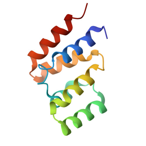
- Primary Citation Related Structures:
8WKW - PubMed Abstract:
Retinoic acid-inducible gene-I (RIG-I)-like receptors (RLRs) and cyclic GMP-AMP synthase (cGAS) recognize aberrant nucleic acids and initiate antiviral responses. Host factor zinc finger CCHC domain-containing protein 3 (ZCCHC3) positively regulates both RLRs- and cGAS-mediated signaling through unknown mechanisms. Here, we show that ZCCHC3 employs a broad and unified strategy to promote these pathways in human cell lines. Rather than developing strong protein-protein interactions, ZCCHC3 harbors multiple nucleic-acid-binding modules and undergoes robust liquid phase condensation with nucleic acids. RNA-induced ZCCHC3 condensates enrich and activate RLRs, which then facilitate the interaction of RLRs with the downstream adaptor mitochondrial antiviral-signaling (MAVS). Direct and high-resolution structure determination of liquid condensates confirms the assembly of active-form MAVS filaments. Furthermore, ZCCHC3 efficiently promotes the condensation and enrichment of DNA, cGAS, ATP, and GTP, thereby enhancing cGAS signaling. ZCCHC3 mutants defective in RNA/DNA-induced condensation lost their regulatory efficiency in both pathways. These results highlight unexpectedly broad connections between biomolecular condensation and innate immunity.
- Science and Technology Innovation Center, Shandong First Medical University & Shandong Academy of Medical Sciences, Jinan 250000, China; National Laboratory of Biomacromolecules, CAS Center for Excellence in Biomacromolecules, Institute of Biophysics, Chinese Academy of Sciences, Beijing 100101, China.
Organizational Affiliation: