8WGK

Crystal structure of Rhodothermus marinus substrate-binding protein (Br soaking)


Experimental Data Snapshot

  • Method: X-RAY DIFFRACTION
  • Resolution: 1.75 Å
  • R-Value Free: 0.261 
  • R-Value Work: 0.222 
  • R-Value Observed: 0.226 

wwPDB Validation   3D Report Full Report


This is version 1.1 of the entry. See complete history


Literature

Structural and bioinformatics analysis of single-domain substrate-binding protein from Rhodothermus marinus.

Nam, K.H.

(2024) Biochem Biophys Rep 37: 101611-101611

  • DOI: https://doi.org/10.1016/j.bbrep.2023.101611
  • Primary Citation of Related Structures:  
    8WGK, 8WGL

  • PubMed Abstract: 

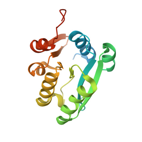
    Substrate-binding proteins (SBPs) are key elements in determining the substrate specificity and high affinity of the ATP-binding cassette uptake system. A typical SBP has two domains that recognize substrates and are responsible for the specific substrate delivery. Conversely, in GenBank, genes for SBPs constituting a single domain SBP are often found in vicinity of a methyl-accepting chemotaxis protein gene. However, the molecular function and mechanism of single domain SBPs are not fully elucidated. To understand their molecular functions, we performed a crystallographic study of single domain SBP from Rhodothermus marinus (RmSBP). RmSBP crystals were soaked in solution containing NaBr or HgCl 2 and their structures determined at 1.75 and 2.3 Å resolution, respectively. RmSBP soaked in NaBr exhibited disorder of the α2-helix, β5-to β6-strand loop, and C-terminus region, showing the structural dynamic region of RmSBP. RmSBP soaked in HgCl 2 showed that Hg 2+ bound to Cys145 located between the α5-and α6-helices. The structural properties of RmSBP were compared with those of single domain SBP homologs. These results will contribute to continued identification of the molecular function and mechanism of single domain SBPs.


  • Organizational Affiliation

    College of General Education, Kookmin University, Seoul, 20707, Republic of Korea.


Macromolecules
Find similar proteins by:  (by identity cutoff)  |  3D Structure
Entity ID: 1
MoleculeChains Sequence LengthOrganismDetailsImage
ABC-type uncharacterized transport system periplasmic component-like protein185Rhodothermus marinus DSM 4252Mutation(s): 0 
Gene Names: Rmar_2176
UniProt
Find proteins for D0MDR1 (Rhodothermus marinus (strain ATCC 43812 / DSM 4252 / R-10))
Explore D0MDR1 
Go to UniProtKB:  D0MDR1
Entity Groups  
Sequence Clusters30% Identity50% Identity70% Identity90% Identity95% Identity100% Identity
UniProt GroupD0MDR1
Sequence Annotations
Expand
  • Reference Sequence
Experimental Data & Validation

Experimental Data

  • Method: X-RAY DIFFRACTION
  • Resolution: 1.75 Å
  • R-Value Free: 0.261 
  • R-Value Work: 0.222 
  • R-Value Observed: 0.226 
  • Space Group: P 21 21 21
Unit Cell:
Length ( Å )Angle ( ˚ )
a = 46.231α = 90
b = 49.129β = 90
c = 57.518γ = 90
Software Package:
Software NamePurpose
PHENIXrefinement
HKL-2000data reduction
HKL-2000data scaling
MOLREPphasing

Structure Validation

View Full Validation Report


Entry History & Funding Information

Deposition Data

  • Released Date: 2023-10-04 
  • Deposition Author(s): Nam, K.H.

Funding OrganizationLocationGrant Number
National Research Foundation (NRF, Korea)Korea, Republic OfNRF-2021R1I1A1A01050838

Revision History  (Full details and data files)

  • Version 1.0: 2023-10-04
    Type: Initial release
  • Version 1.1: 2024-04-17
    Changes: Database references