Membrane structure-responsive lipid scrambling by TMEM63B to control plasma membrane lipid distribution.
Miyata, Y., Takahashi, K., Lee, Y., Sultan, C.S., Kuribayashi, R., Takahashi, M., Hata, K., Bamba, T., Izumi, Y., Liu, K., Uemura, T., Nomura, N., Iwata, S., Nagata, S., Nishizawa, T., Segawa, K.(2025) Nat Struct Mol Biol 32: 185-198
- PubMed: 39424995 Search on PubMedSearch on PubMed Central
- DOI: https://doi.org/10.1038/s41594-024-01411-6
- Primary Citation Related Structures:
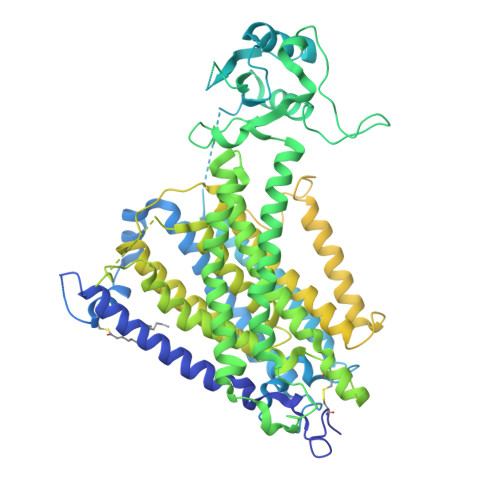
8WG3, 8WG4 - PubMed Abstract:
Phospholipids are asymmetrically distributed in the plasma membrane (PM), with phosphatidylcholine and sphingomyelin abundant in the outer leaflet. However, the mechanisms by which their distribution is regulated remain unclear. Here, we show that transmembrane protein 63B (TMEM63B) functions as a membrane structure-responsive lipid scramblase localized at the PM and lysosomes, activating bidirectional lipid translocation upon changes in membrane curvature and thickness. TMEM63B contains two intracellular loops with palmitoylated cysteine residue clusters essential for its scrambling function. TMEM63B deficiency alters phosphatidylcholine and sphingomyelin distributions in the PM. Persons with heterozygous mutations in TMEM63B are known to develop neurodevelopmental disorders. We show that V44M, the most frequent substitution, confers constitutive scramblase activity on TMEM63B, disrupting PM phospholipid asymmetry. We determined the cryo-electron microscopy structures of TMEM63B in its open and closed conformations, uncovering a lipid translocation pathway formed in response to changes in the membrane environment. Together, our results identify TMEM63B as a membrane structure-responsive scramblase that controls PM lipid distribution and we reveal the molecular basis for lipid scrambling and its biological importance.
- Department of Medical Chemistry, Medical Research Institute, Tokyo Medical and Dental University, Tokyo, Japan.
Organizational Affiliation: