8WDE | pdb_00008wde

CryoEM structure of the spike protein of human CoV 229E in complex with receptor hAPN (composite map)


Experimental Data Snapshot

  • Method: ELECTRON MICROSCOPY
  • Resolution: 3.60 Å
  • Aggregation State: PARTICLE 
  • Reconstruction Method: SINGLE PARTICLE 

wwPDB Validation   3D Report Full Report


This is version 1.1 of the entry. See complete history


Literature

Molecular basis of host recognition of human coronavirus 229E.

Tsai, Y.X.Chien, Y.C.Hsu, M.F.Khoo, K.H.Hsu, S.D.

(2025) Nat Commun 16: 2045-2045

  • DOI: https://doi.org/10.1038/s41467-025-57359-8
  • Primary Citation of Related Structures:  
    8WDE

  • PubMed Abstract: 

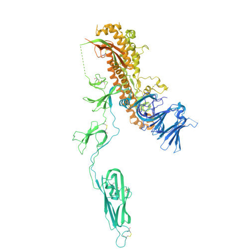
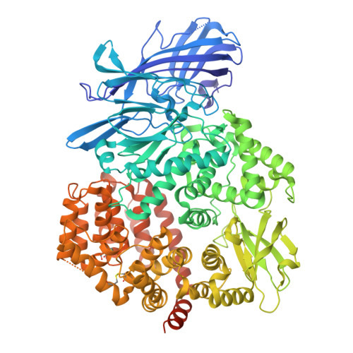
    Human coronavirus 229E (HCoV-229E) is the earliest CoV found to infect humans. It binds to the human aminopeptidase N (hAPN) through the receptor binding domain (RBD) of its spike (S) protein to achieve host recognition. We present the cryo-electron microscopy structure of two HCoV-229E S protein in complex with a dimeric hAPN to provide structural insights on how the HCoV-229E S protein opens up its RBD to engage with its host receptor, information that is currently missing among alphacoronaviruses to which HCoV-229E belong. We quantitatively profile the glycosylation of HCoV-229E S protein and hAPN to deduce the glyco-shielding effects pertinent to antigenicity and host recognition. Finally, we present an atomic model of fully glycosylated HCoV-229E S in complex with hAPN anchored on their respective membrane bilayers to recapitulate the structural basis of the first step of host infection by HCoV-229E.


  • Organizational Affiliation
    • Institute of Biological Chemistry, Academia Sinica, Taipei, 11529, Taiwan.

Macromolecules
Find similar proteins by:  (by identity cutoff)  |  3D Structure
Entity ID: 1
MoleculeChains Sequence LengthOrganismDetailsImage
Spike glycoprotein
A, B, C
1,165Human coronavirus 229EMutation(s): 0 
UniProt
Find proteins for P15423 (Human coronavirus 229E)
Explore P15423 
Go to UniProtKB:  P15423
Entity Groups  
Sequence Clusters30% Identity50% Identity70% Identity90% Identity95% Identity100% Identity
UniProt GroupP15423
Glycosylation
Glycosylation Sites: 8
Sequence Annotations
Expand
  • Reference Sequence
Find similar proteins by:  (by identity cutoff)  |  3D Structure
Entity ID: 2
MoleculeChains Sequence LengthOrganismDetailsImage
Aminopeptidase N
D, E
943Homo sapiensMutation(s): 0 
Gene Names: ANPEP
EC: 3.4.11.2
UniProt & NIH Common Fund Data Resources
Find proteins for P15144 (Homo sapiens)
Explore P15144 
Go to UniProtKB:  P15144
PHAROS:  P15144
GTEx:  ENSG00000166825 
Entity Groups  
Sequence Clusters30% Identity50% Identity70% Identity90% Identity95% Identity100% Identity
UniProt GroupP15144
Glycosylation
Glycosylation Sites: 3Go to GlyGen: P15144-1
Sequence Annotations
Expand
  • Reference Sequence
Oligosaccharides

Help

Entity ID: 3
MoleculeChains Length2D Diagram Glycosylation3D Interactions
beta-D-mannopyranose-(1-4)-2-acetamido-2-deoxy-beta-D-glucopyranose-(1-4)-2-acetamido-2-deoxy-beta-D-glucopyranose
F, H, J
3N-Glycosylation
Glycosylation Resources
GlyTouCan:  G15407YE
GlyCosmos:  G15407YE
GlyGen:  G15407YE
Entity ID: 4
MoleculeChains Length2D Diagram Glycosylation3D Interactions
2-acetamido-2-deoxy-beta-D-glucopyranose-(1-4)-2-acetamido-2-deoxy-beta-D-glucopyranose
G, I, K, L
2N-Glycosylation
Glycosylation Resources
GlyTouCan:  G42666HT
GlyCosmos:  G42666HT
GlyGen:  G42666HT
Small Molecules
Ligands 1 Unique
IDChains Name / Formula / InChI Key2D Diagram3D Interactions
NAG (Subject of Investigation/LOI)
Query on NAG

Download Ideal Coordinates CCD File 
AA [auth C]
BA [auth C]
CA [auth D]
DA [auth D]
EA [auth D]
AA [auth C],
BA [auth C],
CA [auth D],
DA [auth D],
EA [auth D],
FA [auth E],
M [auth A],
N [auth A],
O [auth A],
P [auth A],
Q [auth A],
R [auth B],
S [auth B],
T [auth B],
U [auth B],
V [auth B],
W [auth C],
X [auth C],
Y [auth C],
Z [auth C]
2-acetamido-2-deoxy-beta-D-glucopyranose
C8 H15 N O6
OVRNDRQMDRJTHS-FMDGEEDCSA-N
Experimental Data & Validation

Experimental Data

  • Method: ELECTRON MICROSCOPY
  • Resolution: 3.60 Å
  • Aggregation State: PARTICLE 
  • Reconstruction Method: SINGLE PARTICLE 

Structure Validation

View Full Validation Report


Entry History & Funding Information

Deposition Data


Funding OrganizationLocationGrant Number
Academia Sinica (Taiwan)TaiwanAS-CDA-109-L08
Academia Sinica (Taiwan)TaiwanAS-IDR-112-04
Academia Sinica (Taiwan)TaiwanAS-IDR-111-03
National Science Council (NSC, Taiwan)Taiwan110-2113-M-001-050-MY3
National Science Council (NSC, Taiwan)Taiwan110-2311-B-001-013-MY3
Academia Sinica (Taiwan)TaiwanAS-CFII-111-201
Academia Sinica (Taiwan)TaiwanAS-CFII-111-209
Academia Sinica (Taiwan)TaiwanAS-CFII-111-210
Academia Sinica (Taiwan)TaiwanAS-KPQ-109-TPP2

Revision History  (Full details and data files)

  • Version 1.0: 2025-03-05
    Type: Initial release
  • Version 1.1: 2025-03-12
    Changes: Data collection, Database references