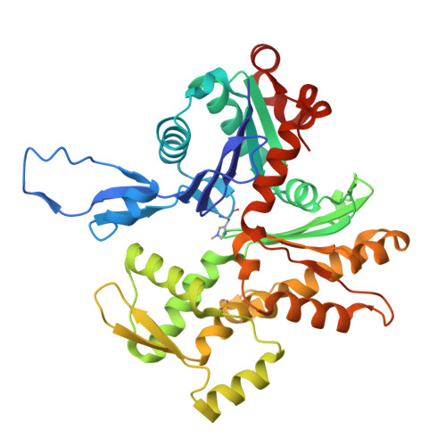
8W36 | pdb_00008w36

rabbit actin in the absence of potassium


Experimental Data Snapshot

  • Method: ELECTRON MICROSCOPY
  • Resolution: 2.27 Å
  • Aggregation State: FILAMENT 
  • Reconstruction Method: HELICAL 

wwPDB Validation 3D Report Full Report

Validation slider image for 8W36

This is version 1.1 of the entry. See complete history

Literature

High-resolution yeast actin structures indicate the molecular mechanism of actin filament stiffening by cations.

Xu, X.P.Cao, W.Swift, M.F.Pandit, N.G.Huehn, A.E.Sindelar, C.V.De La Cruz, E.M.Hanein, D.Volkmann, N.

(2024) Commun Chem 7: 164-164

  • DOI: https://doi.org/10.1038/s42004-024-01243-x
  • Primary Citation Related Structures: 
    8W36

  • PubMed Abstract: 

    Actin filament assembly and the regulation of its mechanical properties are fundamental processes essential for eukaryotic cell function. Residue E167 in vertebrate actins forms an inter-subunit salt bridge with residue K61 of the adjacent subunit. Saccharomyces cerevisiae actin filaments are more flexible than vertebrate filaments and have an alanine at this position (A167). Substitution of this alanine for a glutamic acid (A167E) confers Saccharomyces cerevisiae actin filaments with salt-dependent stiffness similar to vertebrate actins. We developed an optimized cryogenic electron microscopy workflow refining sample preparation and vitrification to obtain near-atomic resolution structures of wild-type and A167E mutant Saccharomyces cerevisiae actin filaments. The difference between these structures allowed us to pinpoint the potential binding site of a filament-associated cation that controls the stiffness of the filaments in vertebrate and A167E Saccharomyces cerevisiae actins. Through an analysis of previously published high-resolution reconstructions of vertebrate actin filaments, along with a newly determined high-resolution vertebrate actin structure in the absence of potassium, we identified a unique peak near residue 167 consistent with the binding of a magnesium ion. Our findings show how magnesium can contribute to filament stiffening by directly bridging actin subunits and allosterically affecting the orientation of the DNase-I binding loop of actin, which plays a regulatory role in modulating actin filament stiffness and interactions with regulatory proteins.


  • Organizational Affiliation
    • Scintillon Institute, 6868 Nancy Ridge Drive, San Diego, CA, 92121, USA.

Macromolecule Content 

  • Total Structure Weight: 209.19 kDa 
  • Atom Count: 14,640 
  • Modeled Residue Count: 1,855 
  • Deposited Residue Count: 1,855 
  • Unique protein chains: 1

Macromolecules

Find similar proteins by:|  3D Structure
Entity ID: 1
MoleculeChains  Sequence LengthOrganismDetailsImage
Actin, alpha skeletal muscleA [auth D],
B [auth C],
C [auth A],
D [auth B],
E
371Oryctolagus cuniculusMutation(s): 0 
Gene Names: ACTA1ACTA
EC: 3.6.4
UniProt
Find proteins for P68135 (Oryctolagus cuniculus)
Explore P68135 
Go to UniProtKB:  P68135
Entity Groups
Sequence Clusters30% Identity50% Identity70% Identity90% Identity95% Identity100% Identity
UniProt GroupP68135
Sequence Annotations
Expand
Reference Sequence

Small Molecules

Ligands 2 Unique
IDChains Name / Formula / InChI Key2D Diagram3D Interactions
ADP
(Subject of Investigation/LOI)

Query on ADP


Download:Ideal Coordinates CCD File
F [auth D],
H [auth C],
J [auth A],
L [auth B],
N [auth E]
ADENOSINE-5'-DIPHOSPHATE
C10 H15 N5 O10 P2
XTWYTFMLZFPYCI-KQYNXXCUSA-N
MG
(Subject of Investigation/LOI)

Query on MG


Download:Ideal Coordinates CCD File
G [auth D],
I [auth C],
K [auth A],
M [auth B],
O [auth E]
MAGNESIUM ION
Mg
JLVVSXFLKOJNIY-UHFFFAOYSA-N
Modified Residues  1 Unique
IDChains TypeFormula2D DiagramParent
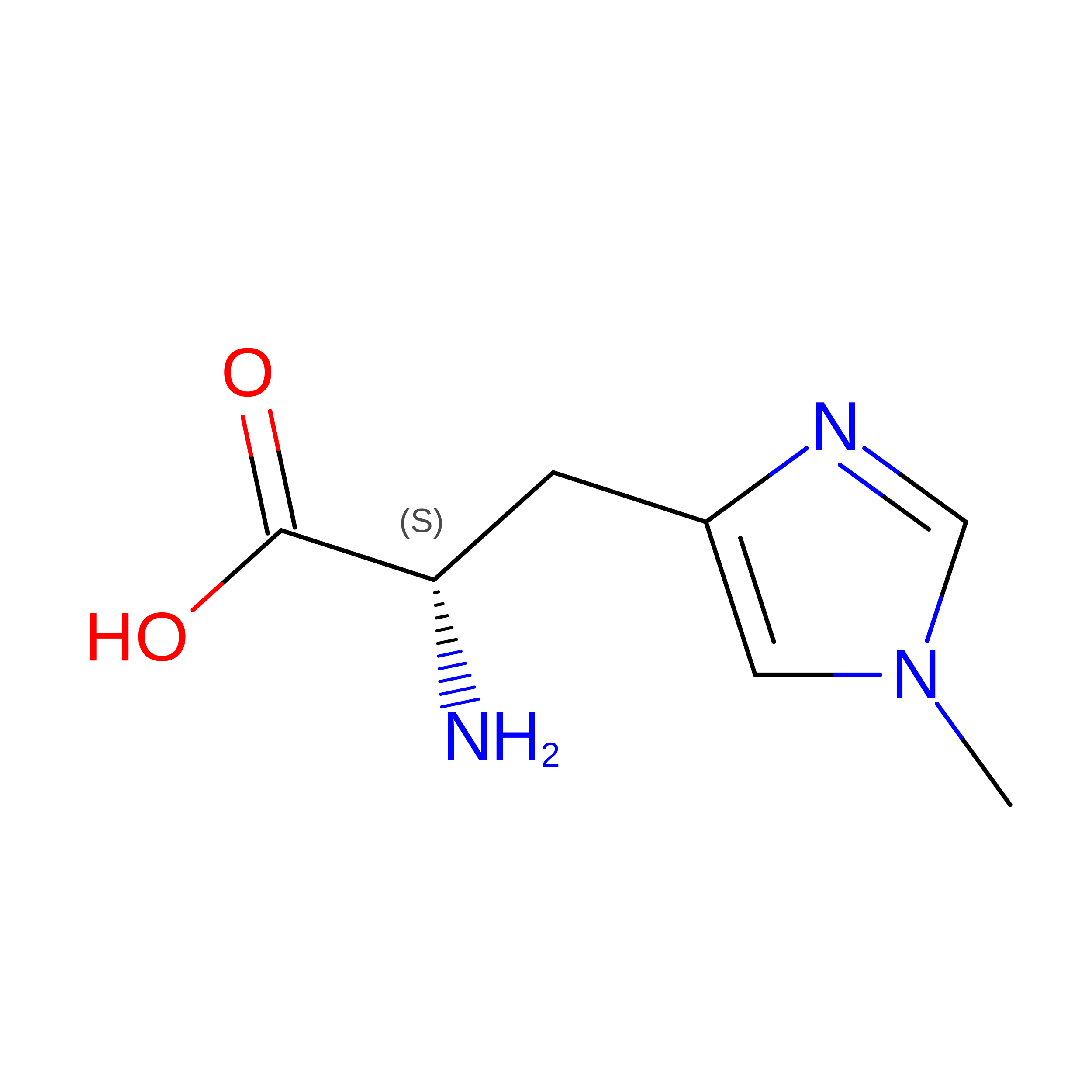
HIC
Query on HIC
A [auth D],
B [auth C],
C [auth A],
D [auth B],
E
L-PEPTIDE LINKINGC7 H11 N3 O2HIS

Experimental Data & Validation

Experimental Data

  • Method: ELECTRON MICROSCOPY
  • Resolution: 2.27 Å
  • Aggregation State: FILAMENT 
  • Reconstruction Method: HELICAL 
EM Software:
TaskSoftware PackageVersion
MODEL REFINEMENTPHENIX1.21_5207:

Structure Validation

View Full Validation Report


Entry History 

& Funding Information

Deposition Data

  • Released Date: 2025-02-26 
  • Deposition Author(s): Volkmann, N.

Funding OrganizationLocationGrant Number
National Institutes of Health/National Institute of General Medical Sciences (NIH/NIGMS)United States--

Revision History  (Full details and data files)

  • Version 1.0: 2025-02-26
    Type: Initial release
  • Version 1.1: 2026-09-16
    Changes: Data collection, Database references