8W1Z | pdb_00008w1z

Structure of a LGR dimer from Caenorhabditis elegans in apo state


Experimental Data Snapshot

  • Method: ELECTRON MICROSCOPY
  • Resolution: 3.79 Å
  • Aggregation State: PARTICLE 
  • Reconstruction Method: SINGLE PARTICLE 

wwPDB Validation   3D Report Full Report


This is version 1.0 of the entry. See complete history


Literature

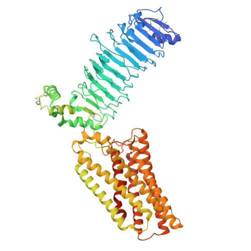
Structure of an LGR dimer, an evolutionary predecessor of glycoprotein hormone receptors.

Gong, Z.Chen, S.Fu, Z.Kloss, B.Wang, C.Kim, J.Clarke, O.B.Fan, Q.R.Hendrickson, W.A.

(2025) Nat Commun 

  • DOI: https://doi.org/10.1038/s41467-025-66676-x
  • Primary Citation of Related Structures:  
    8W1Z

  • PubMed Abstract: 

    Glycoprotein hormones (GpHs) produced in the human pituitary act through receptors (GpHRs) in the gonads to support reproduction and in the thyroid for metabolism. GpHs are heterodimeric cystine-knot proteins; their receptors bind cognate hormones at an extracellular domain and signal through a transmembrane domain to heterotrimeric G proteins. GpHs and GpHRs have co-evolved from invertebrate counterparts. Structures of the human receptors as isolated for cryogenic electron microscopy (cryo-EM) are all monomeric despite compelling evidence for their functioning as dimers. Here we characterize the homologous receptor from Caenorhabditis elegans. Its biochemical properties are notably similar to those of the thyroid stimulating hormone receptor (TSHR) of humans. Structurally, it is an asymmetric dimer (protomers screw-transformed by 142°/4.1 Å), composed such that only one hormone could bind. This is compatible with the 1:2 asymmetry of negatively cooperative TSH:TSHR complexes and for the transactivation evident from functional complementation of binding-deficient and signaling-deficient GpHRs. By modeling, a symmetrized dimer can bind two hormones as in the 2:2 complexes that support TSHR switches in G-protein usage.


  • Organizational Affiliation
    • Department of Biochemistry and Molecular Biophysics, Columbia University, New York, NY, USA.

Macromolecules
Find similar proteins by:  (by identity cutoff)  |  3D Structure
Entity ID: 1
MoleculeChains Sequence LengthOrganismDetailsImage
G-protein coupled receptors family 1 profile domain-containing protein
A, B
701Caenorhabditis elegansMutation(s): 0 
Gene Names: fshr-1lgrC50H2.1CELE_C50H2.1
UniProt
Find proteins for G5EG04 (Caenorhabditis elegans)
Explore G5EG04 
Go to UniProtKB:  G5EG04
Entity Groups  
Sequence Clusters30% Identity50% Identity70% Identity90% Identity95% Identity100% Identity
UniProt GroupG5EG04
Glycosylation
Glycosylation Sites: 3
Sequence Annotations
Expand
  • Reference Sequence
Small Molecules
Ligands 2 Unique
IDChains Name / Formula / InChI Key2D Diagram3D Interactions
CLR
Query on CLR

Download Ideal Coordinates CCD File 
I [auth B]CHOLESTEROL
C27 H46 O
HVYWMOMLDIMFJA-DPAQBDIFSA-N
NAG
Query on NAG

Download Ideal Coordinates CCD File 
C [auth A]
D [auth A]
E [auth A]
F [auth B]
G [auth B]
C [auth A],
D [auth A],
E [auth A],
F [auth B],
G [auth B],
H [auth B]
2-acetamido-2-deoxy-beta-D-glucopyranose
C8 H15 N O6
OVRNDRQMDRJTHS-FMDGEEDCSA-N
Experimental Data & Validation

Experimental Data

  • Method: ELECTRON MICROSCOPY
  • Resolution: 3.79 Å
  • Aggregation State: PARTICLE 
  • Reconstruction Method: SINGLE PARTICLE 
EM Software:
TaskSoftware PackageVersion
MODEL REFINEMENTPHENIX

Structure Validation

View Full Validation Report


Entry History & Funding Information

Deposition Data


Funding OrganizationLocationGrant Number
National Institutes of Health/National Institute of General Medical Sciences (NIH/NIGMS)United StatesNIH P41 GM116799

Revision History  (Full details and data files)

  • Version 1.0: 2025-12-24
    Type: Initial release