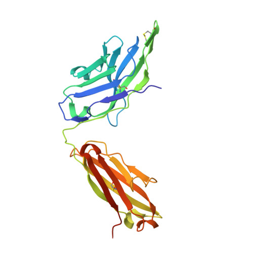
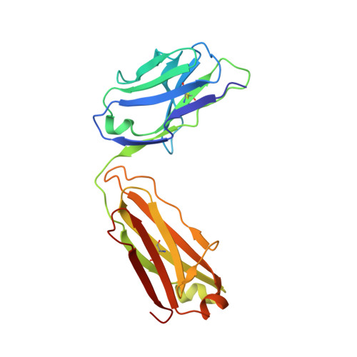
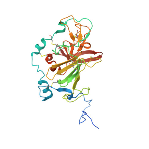
8W0Y

Crystal structure of broadly neutralizing antibody hcab17 in complex with Hepatitis C virus envelope glycoprotein E2 ectodomain


Experimental Data Snapshot

  • Method: X-RAY DIFFRACTION
  • Resolution: 3.31 Å
  • R-Value Free: 0.253 
  • R-Value Work: 0.209 
  • R-Value Observed: 0.211 

wwPDB Validation   3D Report Full Report


Ligand Structure Quality Assessment 


This is version 1.2 of the entry. See complete history


Literature

Convergent evolution and targeting of diverse E2 epitopes by human broadly neutralizing antibodies are associated with HCV clearance.

Ogega, C.O.Skinner, N.E.Schoenle, M.V.Wilcox, X.E.Frumento, N.Wright, D.A.Paul, H.T.Sinnis-Bourozikas, A.Clark, K.E.Figueroa, A.Bjorkman, P.J.Ray, S.C.Flyak, A.I.Bailey, J.R.

(2024) Immunity 57: 890-903.e6

  • DOI: https://doi.org/10.1016/j.immuni.2024.03.001
  • Primary Citation of Related Structures:  
    8W0V, 8W0W, 8W0X, 8W0Y

  • PubMed Abstract: 

    The early appearance of broadly neutralizing antibodies (bNAbs) in serum is associated with spontaneous hepatitis C virus (HCV) clearance, but to date, the majority of bNAbs have been isolated from chronically infected donors. Most of these bNAbs use the V H 1-69 gene segment and target the envelope glycoprotein E2 front layer. Here, we performed longitudinal B cell receptor (BCR) repertoire analysis on an elite neutralizer who spontaneously cleared multiple HCV infections. We isolated 10,680 E2-reactive B cells, performed BCR sequencing, characterized monoclonal B cell cultures, and isolated bNAbs. In contrast to what has been seen in chronically infected donors, the bNAbs used a variety of V H genes and targeted at least three distinct E2 antigenic sites, including sites previously thought to be non-neutralizing. Diverse front-layer-reactive bNAb lineages evolved convergently, acquiring breadth-enhancing somatic mutations. These findings demonstrate that HCV clearance-associated bNAbs are genetically diverse and bind distinct antigenic sites that should be the target of vaccine-induced bNAbs.


  • Organizational Affiliation

    Division of Infectious Diseases, Department of Medicine, Johns Hopkins University School of Medicine, Baltimore, MD, USA; Department of Pharmacology and Molecular Sciences, Johns Hopkins University School of Medicine, Baltimore, MD, USA.


Macromolecules
Find similar proteins by:  (by identity cutoff)  |  3D Structure
Entity ID: 1
MoleculeChains Sequence LengthOrganismDetailsImage
hcab17 Fab Heavy ChainA,
E [auth H]
236Homo sapiensMutation(s): 0 
Entity Groups  
Sequence Clusters30% Identity50% Identity70% Identity90% Identity95% Identity100% Identity
Sequence Annotations
Expand
  • Reference Sequence
Find similar proteins by:  (by identity cutoff)  |  3D Structure
Entity ID: 2
MoleculeChains Sequence LengthOrganismDetailsImage
hcab17 Fab Light ChainB,
F [auth L]
214Homo sapiensMutation(s): 0 
Entity Groups  
Sequence Clusters30% Identity50% Identity70% Identity90% Identity95% Identity100% Identity
Sequence Annotations
Expand
  • Reference Sequence
Find similar proteins by:  (by identity cutoff)  |  3D Structure
Entity ID: 3
MoleculeChains Sequence LengthOrganismDetailsImage
Envelope glycoprotein E2
C, D
262Hepacivirus hominisMutation(s): 0 
UniProt
Find proteins for A0A2P0NE15 (Hepacivirus hominis)
Explore A0A2P0NE15 
Go to UniProtKB:  A0A2P0NE15
Entity Groups  
Sequence Clusters30% Identity50% Identity70% Identity90% Identity95% Identity100% Identity
UniProt GroupA0A2P0NE15
Sequence Annotations
Expand
  • Reference Sequence
Oligosaccharides

Help

Entity ID: 4
MoleculeChains Length2D Diagram Glycosylation3D Interactions
2-acetamido-2-deoxy-beta-D-glucopyranose-(1-4)-2-acetamido-2-deoxy-beta-D-glucopyranose
G [auth E],
H [auth F],
J [auth I],
L [auth K],
M,
G [auth E],
H [auth F],
J [auth I],
L [auth K],
M,
O,
P
2N-Glycosylation
Glycosylation Resources
GlyTouCan:  G42666HT
GlyCosmos:  G42666HT
GlyGen:  G42666HT
Entity ID: 5
MoleculeChains Length2D Diagram Glycosylation3D Interactions
alpha-D-mannopyranose-(1-3)-[alpha-D-mannopyranose-(1-6)]beta-D-mannopyranose-(1-4)-2-acetamido-2-deoxy-beta-D-glucopyranose-(1-4)-2-acetamido-2-deoxy-beta-D-glucopyranoseI [auth G]5N-Glycosylation
Glycosylation Resources
GlyTouCan:  G22768VO
GlyCosmos:  G22768VO
GlyGen:  G22768VO
Entity ID: 6
MoleculeChains Length2D Diagram Glycosylation3D Interactions
beta-D-mannopyranose-(1-4)-2-acetamido-2-deoxy-beta-D-glucopyranose-(1-4)-2-acetamido-2-deoxy-beta-D-glucopyranoseK [auth J],
N
3N-Glycosylation
Glycosylation Resources
GlyTouCan:  G15407YE
GlyCosmos:  G15407YE
GlyGen:  G15407YE
Small Molecules
Ligands 1 Unique
IDChains Name / Formula / InChI Key2D Diagram3D Interactions
NAG
Query on NAG

Download Ideal Coordinates CCD File 
Q [auth C]2-acetamido-2-deoxy-beta-D-glucopyranose
C8 H15 N O6
OVRNDRQMDRJTHS-FMDGEEDCSA-N
Experimental Data & Validation

Experimental Data

  • Method: X-RAY DIFFRACTION
  • Resolution: 3.31 Å
  • R-Value Free: 0.253 
  • R-Value Work: 0.209 
  • R-Value Observed: 0.211 
  • Space Group: P 3 2 1
Unit Cell:
Length ( Å )Angle ( ˚ )
a = 179.41α = 90
b = 179.41β = 90
c = 168.762γ = 120
Software Package:
Software NamePurpose
PHENIXrefinement
Aimlessdata scaling
iMOSFLMdata reduction
PHASERphasing

Structure Validation

View Full Validation Report


Ligand Structure Quality Assessment 


Entry History & Funding Information

Deposition Data


Funding OrganizationLocationGrant Number
National Institutes of Health/National Institute Of Allergy and Infectious Diseases (NIH/NIAID)United StatesU19 AI159822
National Institutes of Health/National Institute Of Allergy and Infectious Diseases (NIH/NIAID)United StatesR00 AI153465
National Institutes of Health/National Institute Of Allergy and Infectious Diseases (NIH/NIAID)United StatesK99 AI153465

Revision History  (Full details and data files)

  • Version 1.0: 2024-03-27
    Type: Initial release
  • Version 1.1: 2024-04-03
    Changes: Database references
  • Version 1.2: 2024-04-17
    Changes: Database references