8VV0 | pdb_00008vv0

CryoEM structure of human GABAA receptor pi (GABRP) in complex with GABA


Experimental Data Snapshot

  • Method: ELECTRON MICROSCOPY
  • Resolution: 3.10 Å
  • Aggregation State: PARTICLE 
  • Reconstruction Method: SINGLE PARTICLE 

wwPDB Validation   3D Report Full Report


This is version 1.2 of the entry. See complete history


Literature

GABA A receptor pi forms channels that stimulate ERK through a G-protein-dependent pathway.

Wang, Y.Zhang, Y.Li, W.Salovska, B.Zhang, J.Li, T.Li, H.Liu, Y.Kaczmarek, L.K.Pusztai, L.Klein, D.E.

(2025) Mol Cell 85: 166-176.e5

  • DOI: https://doi.org/10.1016/j.molcel.2024.11.016
  • Primary Citation of Related Structures:  
    8VSZ, 8VV0

  • PubMed Abstract: 

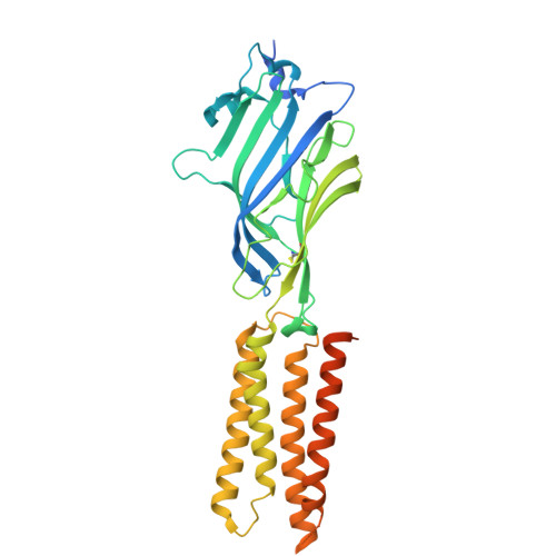
    The rare γ-aminobutyric acid type-A receptor (GABA A R) subunit π (GABRP) is highly expressed in certain cancers, where it stimulates growth through extracellular-regulated kinase (ERK) signaling by an uncharacterized pathway. To elucidate GABRP's signaling mechanism, we determined cryoelectron microscopy (cryo-EM) structures of GABRP embedded in native nanodiscs, both in the presence and absence of GABA. Structurally, GABRP homopentamers closely resemble heteropentameric GABA A R anion channels, transitioning from a closed "resting" state to an open "active" state upon GABA binding. However, functional assays reveal that GABRP responds more like a type-B metabotropic receptor. At physiological concentrations of GABA, chloride flux is not detected. Rather, GABRP activates a G-protein-coupled pathway leading to ERK signaling. Ionotropic activity is only triggered at supraphysiological GABA concentrations, effectively decoupling it from GABRP's signaling functions. These findings provide a structural and functional blueprint for GABRP, opening new avenues for targeted inhibition of GABA growth signals in GABRP-positive cancers.


  • Organizational Affiliation
    • Yale Cancer Biology Institute, Yale University, West Haven, CT 06516, USA; Breast Medical Oncology, Yale Cancer Center, Yale University School of Medicine, New Haven, CT 06520, USA.

Macromolecules
Find similar proteins by:  (by identity cutoff)  |  3D Structure
Entity ID: 1
MoleculeChains Sequence LengthOrganismDetailsImage
Gamma-aminobutyric acid receptor subunit pi
A, B, C, D, E
381Homo sapiensMutation(s): 0 
Gene Names: GABRP
Membrane Entity: Yes 
UniProt & NIH Common Fund Data Resources
Find proteins for O00591 (Homo sapiens)
Explore O00591 
Go to UniProtKB:  O00591
PHAROS:  O00591
GTEx:  ENSG00000094755 
Entity Groups  
Sequence Clusters30% Identity50% Identity70% Identity90% Identity95% Identity100% Identity
UniProt GroupO00591
Glycosylation
Glycosylation Sites: 2Go to GlyGen: O00591-1
Sequence Annotations
Expand
  • Reference Sequence
Oligosaccharides

Help

Entity ID: 2
MoleculeChains Length2D Diagram Glycosylation3D Interactions
alpha-D-mannopyranose-(1-4)-beta-D-mannopyranose-(1-4)-2-acetamido-2-deoxy-beta-D-glucopyranose-(1-4)-2-acetamido-2-deoxy-beta-D-glucopyranose
F, G, H, I, J
4N-Glycosylation
Glycosylation Resources
GlyTouCan:  G08918EL
GlyCosmos:  G08918EL
GlyGen:  G08918EL
Small Molecules
Ligands 2 Unique
IDChains Name / Formula / InChI Key2D Diagram3D Interactions
NAG
Query on NAG

Download Ideal Coordinates CCD File 
K [auth A],
M [auth B],
O [auth C],
R [auth D],
T [auth E]
2-acetamido-2-deoxy-beta-D-glucopyranose
C8 H15 N O6
OVRNDRQMDRJTHS-FMDGEEDCSA-N
ABU (Subject of Investigation/LOI)
Query on ABU

Download Ideal Coordinates CCD File 
L [auth A],
N [auth B],
P [auth C],
Q [auth D],
S [auth E]
GAMMA-AMINO-BUTANOIC ACID
C4 H9 N O2
BTCSSZJGUNDROE-UHFFFAOYSA-N
Experimental Data & Validation

Experimental Data

  • Method: ELECTRON MICROSCOPY
  • Resolution: 3.10 Å
  • Aggregation State: PARTICLE 
  • Reconstruction Method: SINGLE PARTICLE 
EM Software:
TaskSoftware PackageVersion
MODEL REFINEMENTPHENIX

Structure Validation

View Full Validation Report


Entry History & Funding Information

Deposition Data


Funding OrganizationLocationGrant Number
Other private--

Revision History  (Full details and data files)

  • Version 1.0: 2024-12-11
    Type: Initial release
  • Version 1.1: 2025-05-21
    Changes: Data collection
  • Version 1.2: 2026-01-07
    Changes: Data collection, Database references