8VSQ | pdb_00008vsq

Crystal structure of Esub1


Experimental Data Snapshot

  • Method: X-RAY DIFFRACTION
  • Resolution: 2.90 Å
  • R-Value Free: 
    0.281 (Depositor), 0.282 (DCC) 
  • R-Value Work: 
    0.222 (Depositor), 0.224 (DCC) 

Starting Model: in silico
View more details

wwPDB Validation   3D Report Full Report


This is version 1.1 of the entry. See complete history


Literature

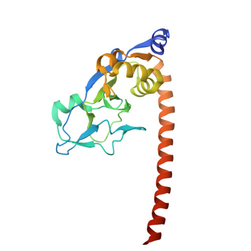
The phage protein paratox is a multifunctional metabolic regulator of Streptococcus.

Muna, T.H.Rutbeek, N.R.Horne, J.Lao, Y.W.Krokhin, O.V.Prehna, G.

(2025) Nucleic Acids Res 53

  • DOI: https://doi.org/10.1093/nar/gkae1200
  • Primary Citation of Related Structures:  
    8VSQ, 8VSR

  • PubMed Abstract: 

    Streptococcus pyogenes, or Group A Streptococcus (GAS), is a commensal bacteria and human pathogen. Central to GAS pathogenesis is the presence of prophage encoded virulence genes. The conserved phage gene for the protein paratox (Prx) is genetically linked to virulence genes, but the reason for this linkage is unknown. Prx inhibits GAS quorum sensing and natural competence by binding the transcription factor ComR. However, inhibiting ComR does not explain the virulence gene linkage. To address this, we took a mass spectrometry approach to search for other Prx interaction partners. The data demonstrates that Prx binds numerous DNA-binding proteins and transcriptional regulators. We show binding of Prx in vitro with the GAS protein Esub1 (SpyM3_0890) and the phage protein JM3 (SpyM3_1246). An Esub1:Prx complex X-ray crystal structure reveals that Esub1 and ComR possess a conserved Prx-binding helix. Computational modelling predicts that the Prx-binding helix is present in several, but not all, binding partners. Namely, JM3 lacks the Prx-binding helix. As Prx is conformationally dynamic, this suggests partner-dependent binding modes. Overall, Prx acts as a metabolic regulator of GAS to maintain the phage genome. As such, Prx maybe a direct contributor to the pathogenic conversion of GAS.


  • Organizational Affiliation

    Department of Microbiology, University of Manitoba, 45 Chancellors Circle, Buller Building, Winnipeg MB, R3T 2N2, Canada.


Macromolecules
Find similar proteins by:  (by identity cutoff)  |  3D Structure
Entity ID: 1
MoleculeChains Sequence LengthOrganismDetailsImage
Esub1
A, B
206Streptococcus pyogenes MGAS315Mutation(s): 0 
Gene Names: SpyM3_0890
UniProt
Find proteins for A0A0H2UUU0 (Streptococcus pyogenes serotype M3 (strain ATCC BAA-595 / MGAS315))
Explore A0A0H2UUU0 
Go to UniProtKB:  A0A0H2UUU0
Entity Groups  
Sequence Clusters30% Identity50% Identity70% Identity90% Identity95% Identity100% Identity
UniProt GroupA0A0H2UUU0
Sequence Annotations
Expand
  • Reference Sequence
Small Molecules
Ligands 1 Unique
IDChains Name / Formula / InChI Key2D Diagram3D Interactions
SO4
Query on SO4

Download Ideal Coordinates CCD File 
C [auth A]SULFATE ION
O4 S
QAOWNCQODCNURD-UHFFFAOYSA-L
Experimental Data & Validation

Experimental Data

  • Method: X-RAY DIFFRACTION
  • Resolution: 2.90 Å
  • R-Value Free:  0.281 (Depositor), 0.282 (DCC) 
  • R-Value Work:  0.222 (Depositor), 0.224 (DCC) 
Space Group: P 1 2 1
Unit Cell:
Length ( Å )Angle ( ˚ )
a = 82.362α = 90
b = 43.872β = 118.668
c = 85.841γ = 90
Software Package:
Software NamePurpose
REFMACrefinement
XDSdata reduction
Aimlessdata scaling
PHENIXphasing

Structure Validation

View Full Validation Report


Entry History & Funding Information

Deposition Data


Funding OrganizationLocationGrant Number
Natural Sciences and Engineering Research Council (NSERC, Canada)CanadaRGPIN-2018-04968

Revision History  (Full details and data files)

  • Version 1.0: 2025-01-08
    Type: Initial release
  • Version 1.1: 2025-01-29
    Changes: Database references