8VRX | pdb_00008vrx

Bile salt hydrolase from Arthrobacter citreus


Experimental Data Snapshot

  • Method: X-RAY DIFFRACTION
  • Resolution: 2.04 Å
  • R-Value Free: 
    0.172 (Depositor), 0.170 (DCC) 
  • R-Value Work: 
    0.133 (Depositor), 0.130 (DCC) 
  • R-Value Observed: 
    0.135 (Depositor) 

Starting Model: experimental
View more details

wwPDB Validation   3D Report Full Report


This is version 1.1 of the entry. See complete history


Literature

Structural and functional analysis of a bile salt hydrolase from the bison microbiome.

Asar, R.Dhindwal, P.Ruzzini, A.

(2024) J Biological Chem 300: 107769-107769

  • DOI: https://doi.org/10.1016/j.jbc.2024.107769
  • Primary Citation of Related Structures:  
    8VRX, 8VSY

  • PubMed Abstract: 

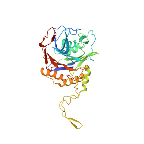
    The bile salt hydrolases (BSHs) are significant constituents of animal microbiomes. An evolving appreciation of their roles in health and disease has established them as targets of pharmacological inhibition. These bacterial enzymes belong to the N-terminal nucleophile superfamily and are best known to catalyze the deconjugation of glycine or taurine from bile salts to release bile acid substrates for transformation and or metabolism in the gastrointestinal tract. Here, we identify and describe the BSH from a common member of the Plains bison microbiome, Arthrobacter citreus (BSH Ac ). Steady-state kinetic analyses demonstrated that BSH Ac is a broad-spectrum hydrolase with a preference for glycine-conjugates and deoxycholic acid (DCA). Second-order rate constants (k cat /K M ) for BSH Ac -catalyzed reactions of relevant bile salts-glyco- and tauro-conjugates of cholic acid and DCA- varied by ∼30-fold and measured between 1.4 × 10 5 and 4.3 × 10 6  M -1 s -1 . Interestingly, a pan-BSH inhibitor named AAA-10 acted as a slow irreversible inhibitor of BSH Ac with a rate of inactivation (k inact ) of ∼2 h -1 and a second order rate constant (k inact /K I ) of ∼24 M -1 s -1 for the process. Structural characterization of BSH Ac reacted with AAA-10 showed covalent modification of the N-terminal cysteine nucleophile, providing molecular details for an enzyme-stabilized product formed from this mechanism-based inhibitor's α-fluoromethyl ketone warhead. Structural comparison of the BSHs and BSH:inhibitor complexes highlighted the plasticity of the steroid-binding site, including a flexible loop that is variable across well-studied BSHs.


  • Organizational Affiliation

    Department of Veterinary Microbiology, Western College of Veterinary Medicine, University of Saskatchewan, Saskatoon, SK, Canada.


Macromolecules
Find similar proteins by:  (by identity cutoff)  |  3D Structure
Entity ID: 1
MoleculeChains Sequence LengthOrganismDetailsImage
Bile Salt Hydrolase
A, B
322Arthrobacter citreusMutation(s): 0 
EC: 3.5.1.24
UniProt
Find proteins for A0AAN0NPC4 (Arthrobacter citreus)
Explore A0AAN0NPC4 
Go to UniProtKB:  A0AAN0NPC4
Entity Groups  
Sequence Clusters30% Identity50% Identity70% Identity90% Identity95% Identity100% Identity
UniProt GroupA0AAN0NPC4
Sequence Annotations
Expand
  • Reference Sequence
Small Molecules
Ligands 5 Unique
IDChains Name / Formula / InChI Key2D Diagram3D Interactions
TRS
Query on TRS

Download Ideal Coordinates CCD File 
G [auth A]2-AMINO-2-HYDROXYMETHYL-PROPANE-1,3-DIOL
C4 H12 N O3
LENZDBCJOHFCAS-UHFFFAOYSA-O
PEG
Query on PEG

Download Ideal Coordinates CCD File 
I [auth A],
J [auth A],
K [auth A],
T [auth B],
X [auth B]
DI(HYDROXYETHYL)ETHER
C4 H10 O3
MTHSVFCYNBDYFN-UHFFFAOYSA-N
GOL
Query on GOL

Download Ideal Coordinates CCD File 
R [auth B],
S [auth B],
V [auth B],
W [auth B]
GLYCEROL
C3 H8 O3
PEDCQBHIVMGVHV-UHFFFAOYSA-N
EDO
Query on EDO

Download Ideal Coordinates CCD File 
D [auth A]
F [auth A]
H [auth A]
L [auth A]
N [auth A]
D [auth A],
F [auth A],
H [auth A],
L [auth A],
N [auth A],
O [auth A],
P [auth A],
U [auth B]
1,2-ETHANEDIOL
C2 H6 O2
LYCAIKOWRPUZTN-UHFFFAOYSA-N
MG
Query on MG

Download Ideal Coordinates CCD File 
C [auth A],
E [auth A],
M [auth A],
Q [auth B]
MAGNESIUM ION
Mg
JLVVSXFLKOJNIY-UHFFFAOYSA-N
Experimental Data & Validation

Experimental Data

  • Method: X-RAY DIFFRACTION
  • Resolution: 2.04 Å
  • R-Value Free:  0.172 (Depositor), 0.170 (DCC) 
  • R-Value Work:  0.133 (Depositor), 0.130 (DCC) 
  • R-Value Observed: 0.135 (Depositor) 
Space Group: C 1 2 1
Unit Cell:
Length ( Å )Angle ( ˚ )
a = 167.787α = 90
b = 44.071β = 111.07
c = 86.592γ = 90
Software Package:
Software NamePurpose
PHENIXrefinement
XDSdata reduction
XSCALEdata scaling
PHASERphasing

Structure Validation

View Full Validation Report


Entry History & Funding Information

Deposition Data


Funding OrganizationLocationGrant Number
Natural Sciences and Engineering Research Council (NSERC, Canada)Canada--

Revision History  (Full details and data files)

  • Version 1.0: 2024-02-07
    Type: Initial release
  • Version 1.1: 2025-04-09
    Changes: Database references, Structure summary