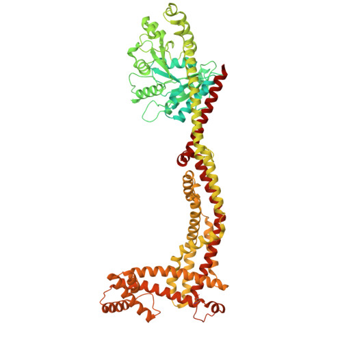
Cardiolipin dynamics promote membrane remodeling by mitochondrial OPA1.
Thatavarthy, S., Abriata, L.A., Meireles, F.T.P., Zuccaro, K.E., Gargey Iragavarapu, A., Sullivan, G.M., Moss 3rd, F.R., Frost, A., Dal Peraro, M., Aydin, H.(2025) Nat Commun 16: 8685-8685
- PubMed: 41027961
- DOI: https://doi.org/10.1038/s41467-025-63813-4
- Primary Citation of Related Structures:
8VLZ, 8VM4 - PubMed Abstract:
Cardiolipin is a mitochondria-specific phospholipid that forms heterotypic interactions with membrane-shaping proteins and regulates the dynamic remodeling and function of mitochondria. However, the precise mechanisms through which cardiolipin influences mitochondrial morphology are not well understood. In this study, employing molecular dynamics simulations, we determined that cardiolipin molecules extensively engage with the paddle domain of mitochondrial fusion protein OPA1, which controls membrane-shaping mechanisms. Structure-function analysis confirmed the interactions between cardiolipin and two conserved motifs of OPA1 at the membrane-binding sites. We further developed a bromine-labeled cardiolipin probe to enhance cryoEM contrast and characterized the structure of OPA1 assemblies bound to the cardiolipin brominated lipid bilayers. Our images provide direct evidence of cardiolipin enrichment within the OPA1-binding leaflet. Last, we observed a decrease in membrane remodeling activity for OPA1 in lipid compositions with increasing concentrations of monolyso-cardiolipin. This suggests that the partial replacement of cardiolipin by monolyso-cardiolipin, as observed in Barth syndrome, alters the malleability of the membrane and compromises proper remodeling. Together, these data provide insights into how biological membranes regulate the mechanisms governing mitochondrial homeostasis.
- Department of Molecular Pathobiology, College of Dentistry, New York University, New York, NY, USA.
Organizational Affiliation: