Identification of the Thermal Activation Network in Human 15-Lipoxygenase-2: Divergence from Plant Orthologs and Its Relationship to Hydrogen Tunneling Activation Barriers.
Ohler, A., Taylor, P.E., Bledsoe, J.A., Iavarone, A.T., Gilbert, N.C., Offenbacher, A.R.(2024) ACS Catal 14: 5444-5457
- PubMed: 38601784 Search on PubMedSearch on PubMed Central
- DOI: https://doi.org/10.1021/acscatal.4c00439
- Primary Citation Related Structures:
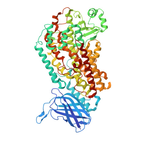
8VIY - PubMed Abstract:
The oxidation of polyunsaturated fatty acids by lipoxygenases (LOXs) is initiated by a C-H cleavage step in which the hydrogen atom is transferred quantum mechanically (i.e., via tunneling). In these reactions, protein thermal motions facilitate the conversion of ground-state enzyme-substrate complexes to tunneling-ready configurations and are thus important for transferring energy from the solvent to the active site for the activation of catalysis. In this report, we employed temperature-dependent hydrogen-deuterium exchange mass spectrometry (TDHDX-MS) to identify catalytically linked, thermally activated peptides in a representative animal LOX, human epithelial 15-LOX-2. TDHDX-MS of wild-type 15-LOX-2 was compared to two active site mutations that retain structural stability but have increased activation energies ( E a ) of catalysis. The E a value of one variant, V427L, is implicated to arise from suboptimal substrate positioning by increased active-site side chain rotamer dynamics, as determined by X-ray crystallography and ensemble refinement. The resolved thermal network from the comparative E a s of TDHDX-MS between wild-type and V426A is localized along the front face of the 15-LOX-2 catalytic domain. The network contains a clustering of isoleucine, leucine, and valine side chains within the helical peptides. This thermal network of 15-LOX-2 is different in location, area, and backbone structure compared to a model plant lipoxygenase from soybean that exhibits a low E a value of catalysis compared to the human ortholog. The presented data provide insights into the divergence of thermally activated protein motions in plant and animal LOXs and their relationships to the enthalpic barriers for facilitating hydrogen tunneling.
- Department of Chemistry, East Carolina University, Greenville, North Carolina 27858, United States.
Organizational Affiliation: