Active site remodeling in tumor-relevant IDH1 mutants drives distinct kinetic features and potential resistance mechanisms.
Mealka, M., Sierra, N.A., Avellaneda Matteo, D., Albekioni, E., Khoury, R., Mai, T., Conley, B.M., Coleman, N.J., Sabo, K.A., Komives, E.A., Bobkov, A.A., Cooksy, A.L., Silletti, S., Schiffer, J.M., Huxford, T., Sohl, C.D.(2024) Nat Commun 15: 3785-3785
- PubMed: 38710674 Search on PubMedSearch on PubMed Central
- DOI: https://doi.org/10.1038/s41467-024-48277-2
- Primary Citation Related Structures:
8VH9, 8VHA, 8VHB, 8VHC, 8VHD, 8VHE - PubMed Abstract:
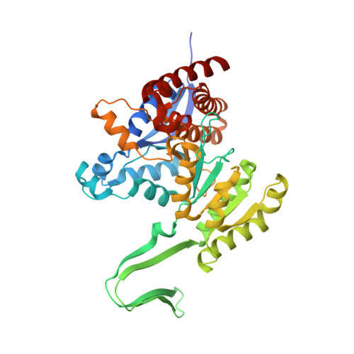
Mutations in human isocitrate dehydrogenase 1 (IDH1) drive tumor formation in a variety of cancers by replacing its conventional activity with a neomorphic activity that generates an oncometabolite. Little is understood of the mechanistic differences among tumor-driving IDH1 mutants. We previously reported that the R132Q mutant unusually preserves conventional activity while catalyzing robust oncometabolite production, allowing an opportunity to compare these reaction mechanisms within a single active site. Here, we employ static and dynamic structural methods and observe that, compared to R132H, the R132Q active site adopts a conformation primed for catalysis with optimized substrate binding and hydride transfer to drive improved conventional and neomorphic activity over R132H. This active site remodeling reveals a possible mechanism of resistance to selective mutant IDH1 therapeutic inhibitors. This work enhances our understanding of fundamental IDH1 mechanisms while pinpointing regions for improving inhibitor selectivity.
- Department of Chemistry & Biochemistry, San Diego State University, San Diego, CA, USA.
Organizational Affiliation: