8VH7 | pdb_00008vh7

Crystal structure of heparosan synthase 2 from Pasteurella multocida at 1.98 A

  • Classification: TRANSFERASE
  • Organism(s): Pasteurella multocida
  • Expression System: Escherichia coli
  • Mutation(s): No 

  • Deposited: 2023-12-31 Released: 2024-07-24 
  • Deposition Author(s): Pedersen, L.C., Liu, J., Stancanelli, E., Krahn, J.M.
  • Funding Organization(s): National Institutes of Health/National Institute of Environmental Health Sciences (NIH/NIEHS), Department of Health & Human Services (HHS), National Institutes of Health/National Heart, Lung, and Blood Institute (NIH/NHLBI)

Experimental Data Snapshot

  • Method: X-RAY DIFFRACTION
  • Resolution: 1.98 Å
  • R-Value Free: 
    0.249 (Depositor), 0.250 (DCC) 
  • R-Value Work: 
    0.208 (Depositor), 0.208 (DCC) 
  • R-Value Observed: 
    0.209 (Depositor) 

Starting Model: experimental
View more details

wwPDB Validation 3D Report Full Report

Validation slider image for 8VH7

Ligand Structure Quality Assessment 


This is version 1.1 of the entry. See complete history

Literature

Structural and Functional Analysis of Heparosan Synthase 2 from Pasteurella multocida (PmHS2) to Improve the Synthesis of Heparin.

Stancanelli, E.Krahn, J.A.Viverette, E.Dutcher, R.Pagadala, V.Borgnia, M.J.Liu, J.Pedersen, L.C.

(2024) ACS Catal 14: 6577-6588

  • DOI: https://doi.org/10.1021/acscatal.4c00677
  • Primary Citation Related Structures: 
    8VH7, 8VH8, 8VIW

  • PubMed Abstract: 

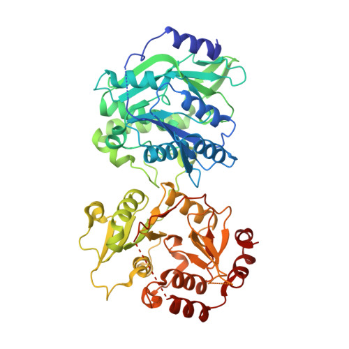
    Heparin is a widely used drug to treat thrombotic disorders in hospitals. Heparosan synthase 2 from Pasteurella multocida (PmHS2) is a key enzyme used for the chemoenzymatic synthesis of heparin oligosaccharides. It has both activities: glucosaminyl transferase activity and glucuronyl transferase activity; however, the mechanism to carry out the glyco-oligomerization is unknown. Here, we report crystal structures of PmHS2 constructs with bound uridine diphosphate (UDP) and a cryo-EM structure of PmHS2 in complex with UDP and a heptasaccharide (NS 7-mer) substrate. Using a LC-MC analytical method, we discovered the enzyme displays both a two-step concerted oligomerization mode and a distributive oligomerization mode depending on the non-reducing end of the starting oligosaccharide primer. Removal of 7 amino acid residues from the C-terminus results in an enzymatically active monomer instead of dimer and loses the concerted oligomerization mode of activity. In addition, the monomer construct can transfer N-acetyl glucosamine at a substrate concentration that is ∼7-fold higher than wildtype enzyme. It was also determined that an F529A mutant can transfer an N-sulfo glucosamine (GlcNS) saccharide from a previously inactive UDP-GlcNS donor. Performing the glyco-transfer reaction at a high substrate concentration and the capability of using unnatural donors are desirable to simplify the chemoenzymatic synthesis to prepare heparin-based therapeutics.


  • Organizational Affiliation
    • Division of Chemical Biology and Medicinal Chemistry, Eshelman School of Pharmacy, University of North Carolina, Chapel Hill, North Carolina, USA.

Macromolecule Content 

  • Total Structure Weight: 130 kDa 
  • Atom Count: 9,307 
  • Modeled Residue Count: 1,044 
  • Deposited Residue Count: 1,104 
  • Unique protein chains: 1

Macromolecules

Find similar proteins by:|  3D Structure
Entity ID: 1
MoleculeChains  Sequence LengthOrganismDetailsImage
Heparosan synthase B
A, B
552Pasteurella multocidaMutation(s): 0 
Gene Names: hssB
UniProt
Find proteins for Q5SGE1 (Pasteurella multocida)
Explore Q5SGE1 
Go to UniProtKB:  Q5SGE1
Entity Groups
Sequence Clusters30% Identity50% Identity70% Identity90% Identity95% Identity100% Identity
UniProt GroupQ5SGE1
Sequence Annotations
Expand
Reference Sequence

Small Molecules

Ligands 4 Unique
IDChains Name / Formula / InChI Key2D Diagram3D Interactions
UDP
(Subject of Investigation/LOI)

Query on UDP


Download:Ideal Coordinates CCD File
D [auth A],
F [auth A],
P [auth B],
R [auth B]
URIDINE-5'-DIPHOSPHATE
C9 H14 N2 O12 P2
XCCTYIAWTASOJW-XVFCMESISA-N
EDO

Query on EDO


Download:Ideal Coordinates CCD File
G [auth A]
H [auth A]
L [auth A]
M [auth A]
N [auth A]
G [auth A],
H [auth A],
L [auth A],
M [auth A],
N [auth A],
T [auth B],
U [auth B],
V [auth B],
W [auth B],
X [auth B],
Y [auth B],
Z [auth B]
1,2-ETHANEDIOL
C2 H6 O2
LYCAIKOWRPUZTN-UHFFFAOYSA-N
MN
(Subject of Investigation/LOI)

Query on MN


Download:Ideal Coordinates CCD File
C [auth A],
E [auth A],
O [auth B],
Q [auth B]
MANGANESE (II) ION
Mn
WAEMQWOKJMHJLA-UHFFFAOYSA-N
NA

Query on NA


Download:Ideal Coordinates CCD File
I [auth A],
J [auth A],
K [auth A],
S [auth B]
SODIUM ION
Na
FKNQFGJONOIPTF-UHFFFAOYSA-N

Experimental Data & Validation

Experimental Data

  • Method: X-RAY DIFFRACTION
  • Resolution: 1.98 Å
  • R-Value Free:  0.249 (Depositor), 0.250 (DCC) 
  • R-Value Work:  0.208 (Depositor), 0.208 (DCC) 
  • R-Value Observed: 0.209 (Depositor) 
Space Group: P 21 21 2
Unit Cell:
Length ( Å )Angle ( ˚ )
a = 93.178α = 90
b = 163.177β = 90
c = 84.273γ = 90
Software Package:
Software NamePurpose
PHENIXrefinement
autoPROCdata reduction
autoPROCdata scaling
PHASERphasing

Structure Validation

View Full Validation Report


Ligand Structure Quality Assessment 


Entry History 

& Funding Information

Deposition Data


Funding OrganizationLocationGrant Number
National Institutes of Health/National Institute of Environmental Health Sciences (NIH/NIEHS)United StatesZIC-ES102645
Department of Health & Human Services (HHS)United StatesR44GM142304
National Institutes of Health/National Heart, Lung, and Blood Institute (NIH/NHLBI)United States1R01HL094463-09

Revision History  (Full details and data files)

  • Version 1.0: 2024-07-24
    Type: Initial release
  • Version 1.1: 2025-03-12
    Changes: Database references, Structure summary