8VH5 | pdb_00008vh5

Cryo-EM structure of Rab12-LRRK2 complex in the LRRK2 dimer state


Experimental Data Snapshot

  • Method: ELECTRON MICROSCOPY
  • Resolution: 4.00 Å
  • Aggregation State: PARTICLE 
  • Reconstruction Method: SINGLE PARTICLE 

wwPDB Validation 3D Report Full Report

Validation slider image for 8VH5

This is version 1.1 of the entry. See complete history. 

Literature

RAB12-LRRK2 complex suppresses primary ciliogenesis and regulates centrosome homeostasis in astrocytes.

Li, X., Zhu, H., Huang, B.T., Li, X., Kim, H., Tan, H., Zhang, Y., Choi, I., Peng, J., Xu, P., Sun, J., Yue, Z.

(2024) Nat Commun 15: 8434-8434

  • DOI: https://doi.org/10.1038/s41467-024-52723-6
  • Primary Citation Related Structures: 
    8VH4, 8VH5

  • PubMed Abstract: 

    The leucine-rich repeat kinase 2 (LRRK2) phosphorylates a subset of RAB GTPases, and their phosphorylation levels are elevated by Parkinson's disease (PD)-linked mutations of LRRK2. However, the precise function of the LRRK2-regulated RAB GTPase in the brain remains to be elucidated. Here, we identify RAB12 as a robust LRRK2 substrate in the mouse brain through phosphoproteomics profiling and solve the structure of RAB12-LRRK2 protein complex through Cryo-EM analysis. Mechanistically, RAB12 cooperates with LRRK2 to inhibit primary ciliogenesis and regulate centrosome homeostasis in astrocytes through enhancing the phosphorylation of RAB10 and recruiting RILPL1, while the functions of RAB12 require a direct interaction with LRRK2 and LRRK2 activity. Furthermore, the ciliary and centrosome defects caused by the PD-linked LRRK2-G2019S mutation are prevented by Rab12 deletion in astrocytes. Thus, our study reveals a physiological function of the RAB12-LRRK2 complex in regulating ciliogenesis and centrosome homeostasis. The RAB12-LRRK2 structure offers a guidance in the therapeutic development of PD by targeting the RAB12-LRRK2 interaction.


  • Organizational Affiliation: 
    • Department of Neurology, Friedman Brain Institute, Icahn School of Medicine at Mount Sinai, New York, NY, USA.

Macromolecule Content 

  • Total Structure Weight: 616.26 kDa 
  • Atom Count: 33,744 
  • Modeled Residue Count: 4,784 
  • Deposited Residue Count: 5,406 
  • Unique protein chains: 2

Macromolecules

Find similar proteins by:|  3D Structure
Entity ID: 1
MoleculeChains  Sequence LengthOrganismDetailsImage
Leucine-rich repeat serine/threonine-protein kinase 2
A, C
2,527Homo sapiensMutation(s): 0 
Gene Names: LRRK2, PARK8
EC: 2.7.11.1 (PDB Primary Data), 3.6.5 (PDB Primary Data)
UniProt & NIH Common Fund Data Resources
Find proteins for Q5S007 (Homo sapiens)
Explore Q5S007 
Go to UniProtKB:  Q5S007
PHAROS:  Q5S007
GTEx:  ENSG00000188906 
Entity Groups
Sequence Clusters30% Identity50% Identity70% Identity90% Identity95% Identity100% Identity
UniProt GroupQ5S007
Sequence Annotations
Expand
Reference Sequence
Find similar proteins by:|  3D Structure
Entity ID: 2
MoleculeChains  Sequence LengthOrganismDetailsImage
Ras-related protein Rab-12
B, D
176Homo sapiensMutation(s): 0 
Gene Names: RAB12
EC: 3.6.5.2
UniProt & NIH Common Fund Data Resources
Find proteins for Q6IQ22 (Homo sapiens)
Explore Q6IQ22 
Go to UniProtKB:  Q6IQ22
PHAROS:  Q6IQ22
GTEx:  ENSG00000206418 
Entity Groups
Sequence Clusters30% Identity50% Identity70% Identity90% Identity95% Identity100% Identity
UniProt GroupQ6IQ22
Sequence Annotations
Expand
Reference Sequence

Small Molecules

Ligands 4 Unique
IDChains Name / Formula / InChI Key2D Diagram3D Interactions
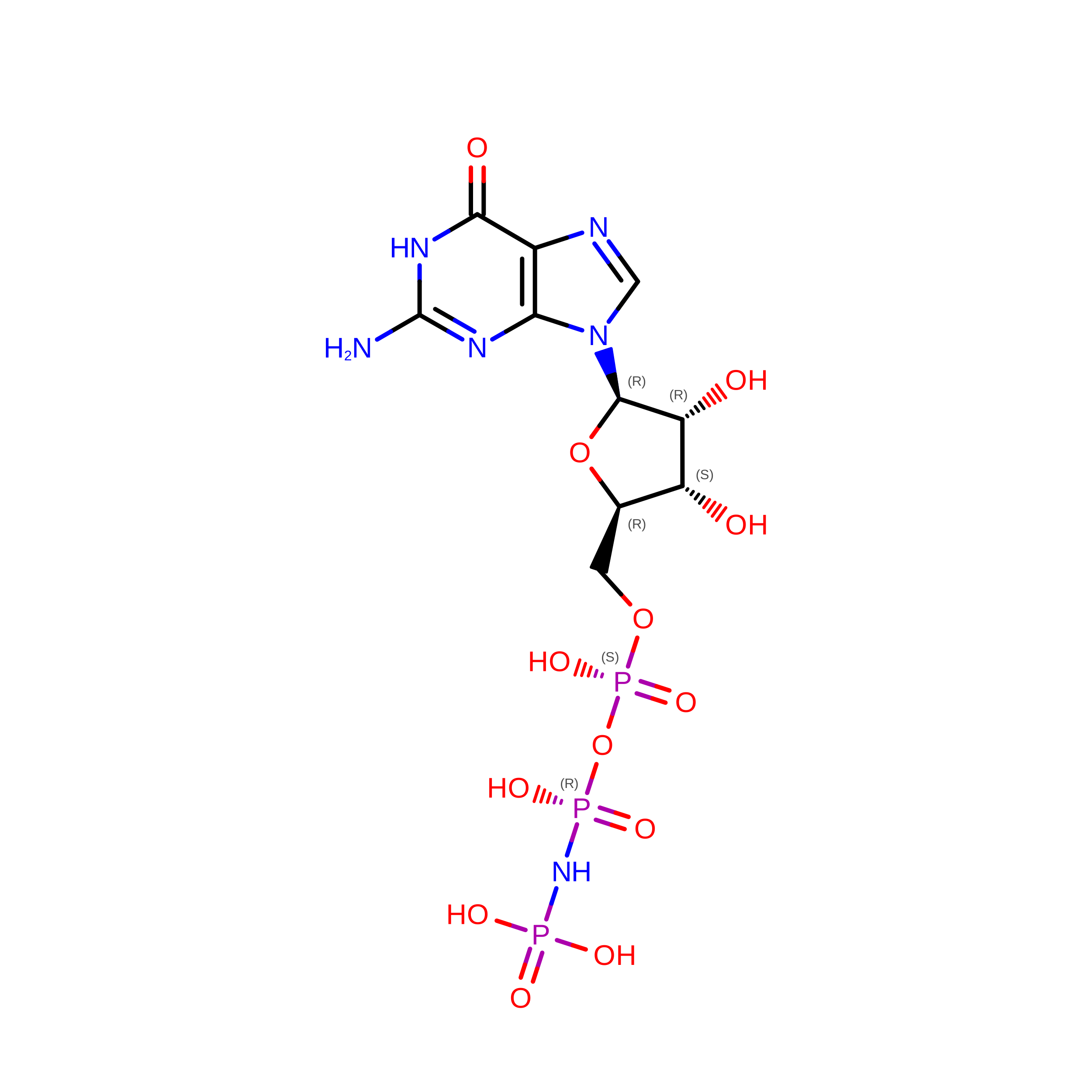
GNP
(Subject of Investigation/LOI)

Query on GNP


Download:Ideal Coordinates CCD File
H [auth B],
L [auth D]
PHOSPHOAMINOPHOSPHONIC ACID-GUANYLATE ESTER
C10 H17 N6 O13 P3
UQABYHGXWYXDTK-UUOKFMHZSA-N
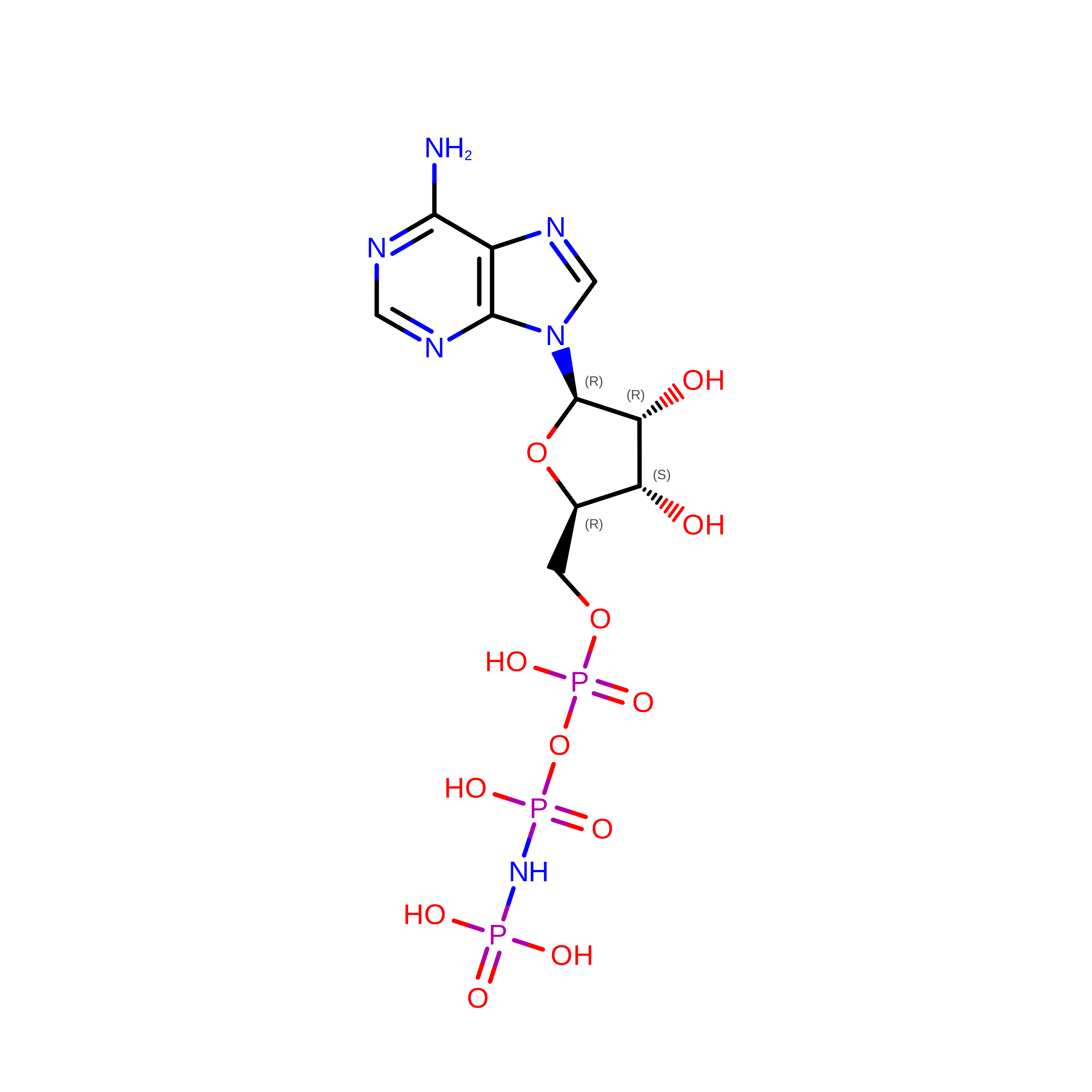
ANP

Query on ANP


Download:Ideal Coordinates CCD File
F [auth A],
J [auth C]
PHOSPHOAMINOPHOSPHONIC ACID-ADENYLATE ESTER
C10 H17 N6 O12 P3
PVKSNHVPLWYQGJ-KQYNXXCUSA-N
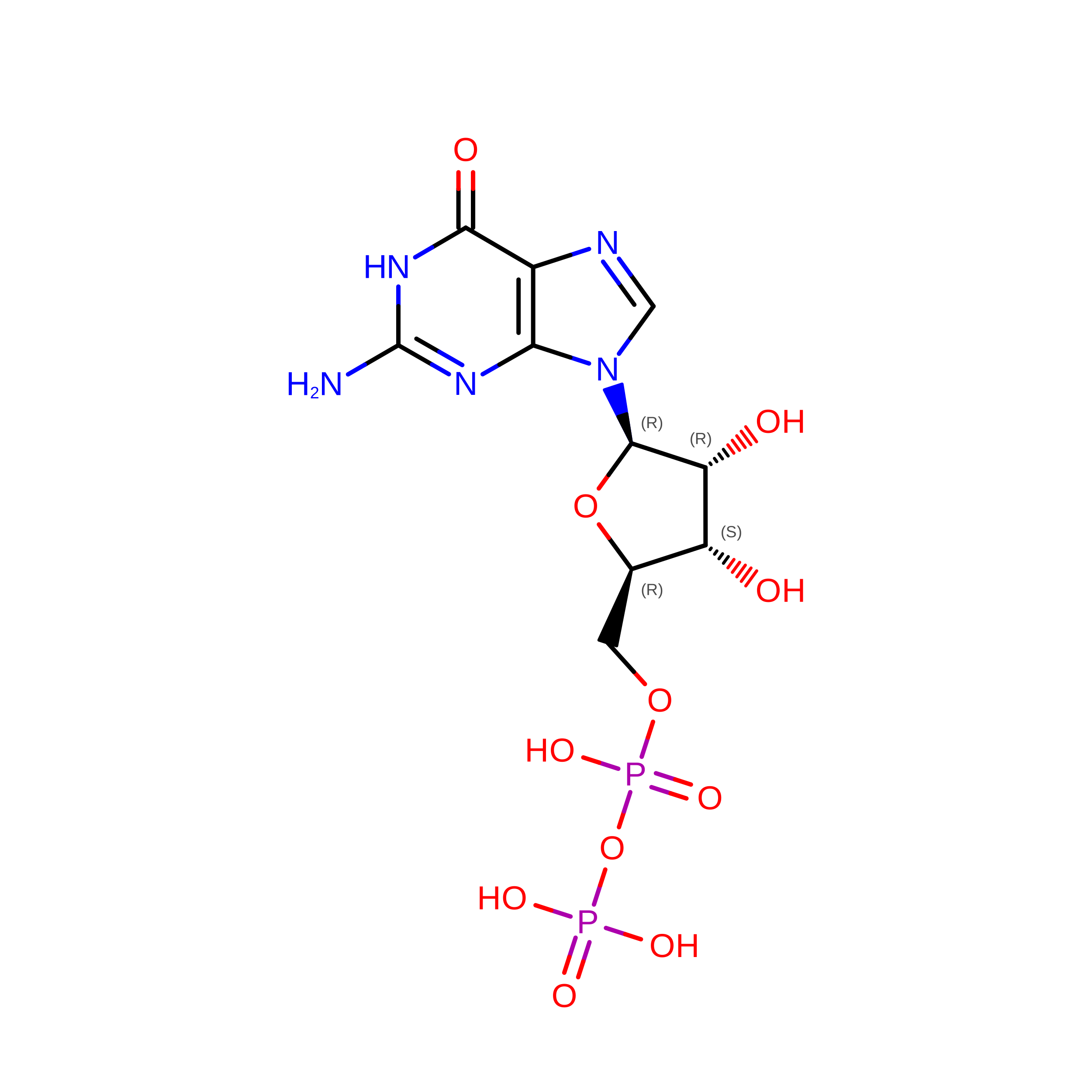
GDP

Query on GDP


Download:Ideal Coordinates CCD File
E [auth A],
I [auth C]
GUANOSINE-5'-DIPHOSPHATE
C10 H15 N5 O11 P2
QGWNDRXFNXRZMB-UUOKFMHZSA-N
MG

Query on MG


Download:Ideal Coordinates CCD File
G [auth B],
K [auth D]
MAGNESIUM ION
Mg
JLVVSXFLKOJNIY-UHFFFAOYSA-N

Experimental Data & Validation

Experimental Data

  • Method: ELECTRON MICROSCOPY
  • Resolution: 4.00 Å
  • Aggregation State: PARTICLE 
  • Reconstruction Method: SINGLE PARTICLE 
EM Software:
TaskSoftware PackageVersion
MODEL REFINEMENTPHENIX
RECONSTRUCTIONcryoSPARC

Structure Validation

View Full Validation Report


Entry History 

& Funding Information

Deposition Data


Funding OrganizationLocationGrant Number
National Institutes of Health/National Cancer Institute (NIH/NCI)United StatesR01NS129795

Revision History  (Full details and data files)

  • Version 1.0: 2024-10-09
    Type: Initial release
  • Version 1.1: 2025-05-28
    Changes: Data collection