8VDW

X-Ray Crystal Structure of the biotin synthase from V. parvula


Experimental Data Snapshot

  • Method: X-RAY DIFFRACTION
  • Resolution: 1.81 Å
  • R-Value Free: 0.227 
  • R-Value Work: 0.179 
  • R-Value Observed: 0.180 

wwPDB Validation   3D Report Full Report


Ligand Structure Quality Assessment 


This is version 1.1 of the entry. See complete history


Literature

Discovery of a Biotin Synthase That Utilizes an Auxiliary 4Fe-5S Cluster for Sulfur Insertion.

Lachowicz, J.C.Lennox-Hvenekilde, D.Myling-Petersen, N.Salomonsen, B.Verkleij, G.Acevedo-Rocha, C.G.Caddell, B.Gronenberg, L.S.Almo, S.C.Sommer, M.O.A.Genee, H.J.Grove, T.L.

(2024) J Am Chem Soc 146: 1860-1873

  • DOI: https://doi.org/10.1021/jacs.3c05481
  • Primary Citation of Related Structures:  
    8VCW, 8VDW

  • PubMed Abstract: 

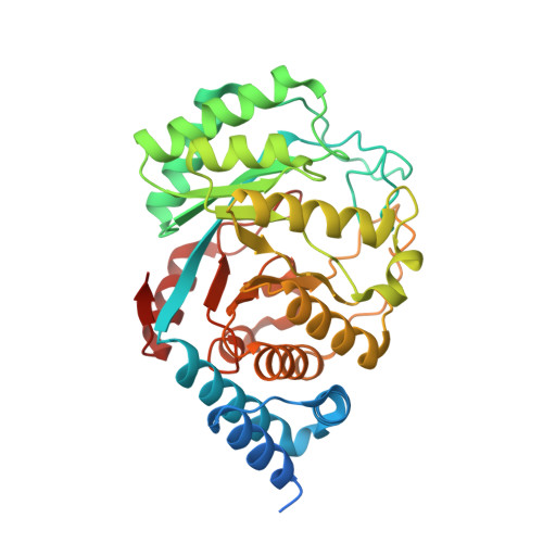
    Biotin synthase (BioB) is a member of the Radical SAM superfamily of enzymes that catalyzes the terminal step of biotin (vitamin B7) biosynthesis, in which it inserts a sulfur atom in desthiobiotin to form a thiolane ring. How BioB accomplishes this difficult reaction has been the subject of much controversy, mainly around the source of the sulfur atom. However, it is now widely accepted that the sulfur atom inserted to form biotin stems from the sacrifice of the auxiliary 2Fe-2S cluster of BioB. Here, we bioinformatically explore the diversity of BioBs available in sequence databases and find an unexpected variation in the coordination of the auxiliary iron-sulfur cluster. After in vitro characterization, including the determination of biotin formation and representative crystal structures, we report a new type of BioB utilized by virtually all obligate anaerobic organisms. Instead of a 2Fe-2S cluster, this novel type of BioB utilizes an auxiliary 4Fe-5S cluster. Interestingly, this auxiliary 4Fe-5S cluster contains a ligated sulfide that we propose is used for biotin formation. We have termed this novel type of BioB, Type II BioB, with the E. coli 2Fe-2S cluster sacrificial BioB representing Type I. This surprisingly ubiquitous Type II BioB has implications for our understanding of the function and evolution of Fe-S clusters in enzyme catalysis, highlighting the difference in strategies between the anaerobic and aerobic world.


  • Organizational Affiliation

    Department of Biochemistry, Albert Einstein College of Medicine, Bronx, New York 10461, United States.


Macromolecules
Find similar proteins by:  (by identity cutoff)  |  3D Structure
Entity ID: 1
MoleculeChains Sequence LengthOrganismDetailsImage
Biotin synthase
A, B
364Veillonella parvulaMutation(s): 0 
Gene Names: bioBHSIVP1_982
EC: 2.8.1.6
UniProt
Find proteins for A0A134C0W5 (Veillonella parvula)
Explore A0A134C0W5 
Go to UniProtKB:  A0A134C0W5
Entity Groups  
Sequence Clusters30% Identity50% Identity70% Identity90% Identity95% Identity100% Identity
UniProt GroupA0A134C0W5
Sequence Annotations
Expand
  • Reference Sequence
Small Molecules
Ligands 5 Unique
IDChains Name / Formula / InChI Key2D Diagram3D Interactions
SAM (Subject of Investigation/LOI)
Query on SAM

Download Ideal Coordinates CCD File 
D [auth A],
K [auth B]
S-ADENOSYLMETHIONINE
C15 H22 N6 O5 S
MEFKEPWMEQBLKI-FCKMPRQPSA-N
Q46 (Subject of Investigation/LOI)
Query on Q46

Download Ideal Coordinates CCD File 
C [auth A],
H [auth B]
Fe4 H S5
Fe4 H S5
IBBAXWHZAAYJDG-UHFFFAOYSA-M
SF4 (Subject of Investigation/LOI)
Query on SF4

Download Ideal Coordinates CCD File 
E [auth A],
J [auth B]
IRON/SULFUR CLUSTER
Fe4 S4
LJBDFODJNLIPKO-UHFFFAOYSA-N
DTB (Subject of Investigation/LOI)
Query on DTB

Download Ideal Coordinates CCD File 
F [auth A],
I [auth B]
6-(5-METHYL-2-OXO-IMIDAZOLIDIN-4-YL)-HEXANOIC ACID
C10 H18 N2 O3
AUTOLBMXDDTRRT-JGVFFNPUSA-N
NA
Query on NA

Download Ideal Coordinates CCD File 
G [auth A]SODIUM ION
Na
FKNQFGJONOIPTF-UHFFFAOYSA-N
Experimental Data & Validation

Experimental Data

  • Method: X-RAY DIFFRACTION
  • Resolution: 1.81 Å
  • R-Value Free: 0.227 
  • R-Value Work: 0.179 
  • R-Value Observed: 0.180 
  • Space Group: P 21 21 21
Unit Cell:
Length ( Å )Angle ( ˚ )
a = 47.465α = 90
b = 70.218β = 90
c = 195.85γ = 90
Software Package:
Software NamePurpose
REFMACrefinement
XDSdata reduction
SCALAdata scaling
PHASERphasing

Structure Validation

View Full Validation Report


Ligand Structure Quality Assessment 


Entry History & Funding Information

Deposition Data


Funding OrganizationLocationGrant Number
National Institutes of Health/National Institute of General Medical Sciences (NIH/NIGMS)United StatesT32 GM007491

Revision History  (Full details and data files)

  • Version 1.0: 2024-01-24
    Type: Initial release
  • Version 1.1: 2024-02-07
    Changes: Database references