Generalized biomolecular modeling and design with RoseTTAFold All-Atom.
Krishna, R., Wang, J., Ahern, W., Sturmfels, P., Venkatesh, P., Kalvet, I., Lee, G.R., Morey-Burrows, F.S., Anishchenko, I., Humphreys, I.R., McHugh, R., Vafeados, D., Li, X., Sutherland, G.A., Hitchcock, A., Hunter, C.N., Kang, A., Brackenbrough, E., Bera, A.K., Baek, M., DiMaio, F., Baker, D.(2024) Science 384: eadl2528-eadl2528
- PubMed: 38452047 Search on PubMed
- DOI: https://doi.org/10.1126/science.adl2528
- Primary Citation Related Structures:
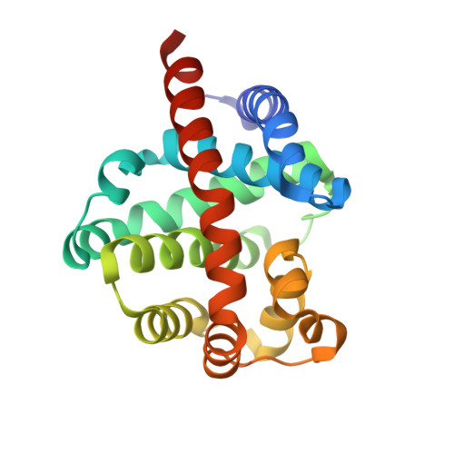
8VC8 - PubMed Abstract:
Deep-learning methods have revolutionized protein structure prediction and design but are presently limited to protein-only systems. We describe RoseTTAFold All-Atom (RFAA), which combines a residue-based representation of amino acids and DNA bases with an atomic representation of all other groups to model assemblies that contain proteins, nucleic acids, small molecules, metals, and covalent modifications, given their sequences and chemical structures. By fine-tuning on denoising tasks, we developed RFdiffusion All-Atom (RFdiffusionAA), which builds protein structures around small molecules. Starting from random distributions of amino acid residues surrounding target small molecules, we designed and experimentally validated, through crystallography and binding measurements, proteins that bind the cardiac disease therapeutic digoxigenin, the enzymatic cofactor heme, and the light-harvesting molecule bilin.
- Department of Biochemistry, University of Washington, Seattle, WA 98105, USA.
Organizational Affiliation: