8V9O | pdb_00008v9o

Imaging scaffold engineered to bind the therapeutic protein target BARD1


Experimental Data Snapshot

  • Method: X-RAY DIFFRACTION
  • Resolution: 3.81 Å
  • R-Value Free: 
    0.231 (Depositor), 0.231 (DCC) 
  • R-Value Work: 
    0.197 (Depositor), 0.197 (DCC) 
  • R-Value Observed: 
    0.200 (Depositor) 

Starting Model: experimental
View more details

wwPDB Validation 3D Report Full Report

Validation slider image for 8V9O

This is version 1.2 of the entry. See complete history

Literature

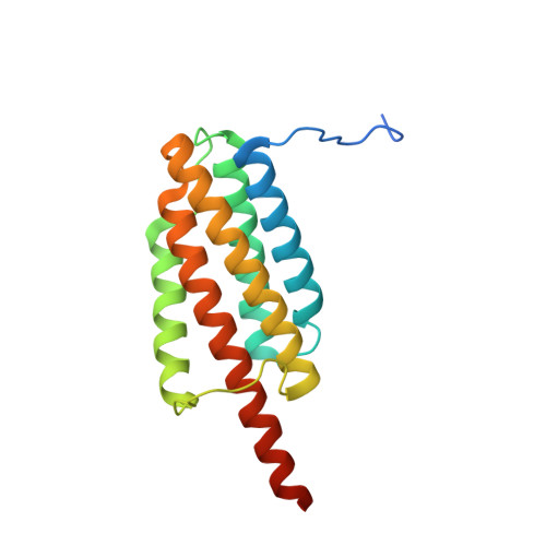
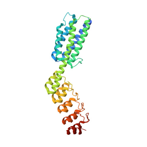
X-ray crystal structure of a designed rigidified imaging scaffold in the ligand-free conformation.

Agdanowski, M.P.Castells-Graells, R.Sawaya, M.R.Cascio, D.Yeates, T.O.Arbing, M.A.

(2024) Acta Crystallogr F Struct Biol Commun 80: 107-115

  • DOI: https://doi.org/10.1107/S2053230X2400414X
  • Primary Citation Related Structures: 
    8V9O

  • PubMed Abstract: 

    Imaging scaffolds composed of designed protein cages fused to designed ankyrin repeat proteins (DARPins) have enabled the structure determination of small proteins by cryogenic electron microscopy (cryo-EM). One particularly well characterized scaffold type is a symmetric tetrahedral assembly composed of 24 subunits, 12 A and 12 B, which has three cargo-binding DARPins positioned on each vertex. Here, the X-ray crystal structure of a representative tetrahedral scaffold in the apo state is reported at 3.8 Å resolution. The X-ray crystal structure complements recent cryo-EM findings on a closely related scaffold, while also suggesting potential utility for crystallographic investigations. As observed in this crystal structure, one of the three DARPins, which serve as modular adaptors for binding diverse `cargo' proteins, present on each of the vertices is oriented towards a large solvent channel. The crystal lattice is unusually porous, suggesting that it may be possible to soak crystals of the scaffold with small (≤30 kDa) protein cargo ligands and subsequently determine cage-cargo structures via X-ray crystallography. The results suggest the possibility that cryo-EM scaffolds may be repurposed for structure determination by X-ray crystallography, thus extending the utility of electron-microscopy scaffold designs for alternative structural biology applications.


  • Organizational Affiliation
    • Department of Chemistry and Biochemistry, University of California Los Angeles, Los Angeles, CA 90095, USA.

Macromolecule Content 

  • Total Structure Weight: 165.8 kDa 
  • Atom Count: 10,640 
  • Modeled Residue Count: 1,374 
  • Deposited Residue Count: 1,500 
  • Unique protein chains: 2

Macromolecules

Find similar proteins by:|  3D Structure
Entity ID: 1
MoleculeChains  Sequence LengthOrganismDetailsImage
Tetrahedral Nanocage Cage, Non-Fusion Component
A, B, C
178synthetic constructMutation(s): 0 
Find similar proteins by:|  3D Structure
Entity ID: 2
MoleculeChains  Sequence LengthOrganismDetailsImage
Tetrahedral Nanocage Cage Component Fused to Anti-BARD1 Darpin
D, E, F
322synthetic constructMutation(s): 0 

Small Molecules

Ligands 1 Unique
IDChains Name / Formula / InChI Key2D Diagram3D Interactions
CA

Query on CA


Download:Ideal Coordinates CCD File
G [auth A]CALCIUM ION
Ca
BHPQYMZQTOCNFJ-UHFFFAOYSA-N

Experimental Data & Validation

Experimental Data

  • Method: X-RAY DIFFRACTION
  • Resolution: 3.81 Å
  • R-Value Free:  0.231 (Depositor), 0.231 (DCC) 
  • R-Value Work:  0.197 (Depositor), 0.197 (DCC) 
  • R-Value Observed: 0.200 (Depositor) 
Space Group: I 2 2 2
Unit Cell:
Length ( Å )Angle ( ˚ )
a = 128.01α = 90
b = 195.57β = 90
c = 228.37γ = 90
Software Package:
Software NamePurpose
PHENIXrefinement
XSCALEdata scaling
XDSdata reduction
PHASERphasing

Structure Validation

View Full Validation Report


Entry History 

& Funding Information

Deposition Data


Funding OrganizationLocationGrant Number
National Institutes of Health/National Institute of General Medical Sciences (NIH/NIGMS)United StatesR01GM129854
Department of Energy (DOE, United States)United StatesDE-FC02-02ER63421

Revision History  (Full details and data files)

  • Version 1.0: 2024-05-15
    Type: Initial release
  • Version 1.1: 2024-06-05
    Changes: Database references
  • Version 1.2: 2024-06-12
    Changes: Database references