Water-directed pinning is key to tau prion formation.
Vigers, M.P., Lobo, S., Najafi, S., Dubose, A., Tsay, K., Ganguly, P., Longhini, A.P., Jin, Y., Buratto, S.K., Kosik, K.S., Shell, M.S., Shea, J.E., Han, S.(2025) Proc Natl Acad Sci U S A 122: e2421391122-e2421391122
- PubMed: 40294272 Search on PubMedSearch on PubMed Central
- DOI: https://doi.org/10.1073/pnas.2421391122
- Primary Citation Related Structures:
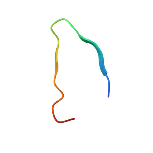
8V1N - PubMed Abstract:
Tau forms fibrillar aggregates that are pathological hallmarks of a family of neurodegenerative diseases known as tauopathies. The synthetic replication of disease-specific fibril structures is a critical gap for developing diagnostic and therapeutic tools. This study debuts a strategy of identifying a critical and minimal folding motif in fibrils characteristic of tauopathies and generating seeding-competent fibrils from the isolated tau peptides. The 19-residue jR2R3 peptide (295 to 313) which spans the R2/R3 splice junction of tau, and includes the P301L mutation, is one such peptide that forms prion-competent fibrils. This tau fragment contains the hydrophobic VQIVYK hexapeptide that is part of the core of all known pathological tau fibril structures and an intramolecular counterstrand that stabilizes the strand-loop-strand (SLS) motif observed in 4R tauopathy fibrils. This study shows that P301L exhibits a duality of effects: it lowers the barrier for the peptide to adopt aggregation-prone conformations and enhances the local structuring of water around the mutation site to facilitate site-directed pinning and dewetting around sites 300-301 to achieve in-register stacking of tau to cross β-sheets. We solved a 3 Å cryo-EM structure of jR2R3-P301L fibrils in which each protofilament layer contains two jR2R3-P301L copies, of which one adopts a SLS fold found in 4R tauopathies and the other wraps around the SLS fold to stabilize it, reminiscent of the three- and fourfold structures observed in 4R tauopathies. These jR2R3-P301L fibrils are competent to template full-length 4R tau in a prion-like manner.
- Department of Chemistry and Biochemistry, University of California, Santa Barbara, CA 93106.
Organizational Affiliation: