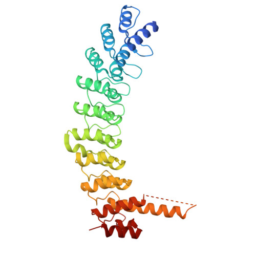
8V0E

ANK repeat of MIB1

  • Classification: LIGASE
  • Organism(s): Homo sapiens
  • Expression System: Escherichia coli
  • Mutation(s): No 

  • Deposited: 2023-11-17 Released: 2024-09-25 
  • Deposition Author(s): Cao, R., Blacklow, S.C.
  • Funding Organization(s): National Institutes of Health/National Cancer Institute (NIH/NCI)

Experimental Data Snapshot

  • Method: X-RAY DIFFRACTION
  • Resolution: 2.39 Å
  • R-Value Free: 0.244 
  • R-Value Work: 0.207 
  • R-Value Observed: 0.208 

Starting Model: experimental
View more details

wwPDB Validation   3D Report Full Report


This is version 1.0 of the entry. See complete history


Literature

Structural requirements for activity of Mind bomb1 in Notch signaling.

Cao, R.Gozlan, O.Airich, A.Tveriakhina, L.Zhou, H.Jiang, H.Cole, P.A.Aster, J.C.Klein, T.Sprinzak, D.Blacklow, S.C.

(2024) Structure 

  • DOI: https://doi.org/10.1016/j.str.2024.07.011
  • Primary Citation of Related Structures:  
    8V0E

  • PubMed Abstract: 

    Mind bomb 1 (MIB1) is a RING E3 ligase that ubiquitinates Notch ligands, a necessary step for induction of Notch signaling. The structural basis for binding of the JAG1 ligand by the N-terminal region of MIB1 is known, yet how the ankyrin (ANK) and RING domains of MIB1 cooperate to catalyze ubiquitin transfer from E2∼Ub to Notch ligands remains unclear. Here, we show that the third RING domain and adjacent coiled coil region (ccRING3) drive MIB1 dimerization and that MIB1 ubiquitin transfer activity relies solely on ccRING3. We report X-ray crystal structures of a UbcH5B-ccRING3 complex and the ANK domain. Directly tethering the MIB1 N-terminal region to ccRING3 forms a minimal MIB1 protein sufficient to induce a Notch response in receiver cells and rescue mib knockout phenotypes in flies. Together, these studies define the functional elements of an E3 ligase needed for ligands to induce a Notch signaling response.


  • Organizational Affiliation

    Department of Biological Chemistry and Molecular Pharmacology, Blavatnik Institute, Harvard Medical School, Boston, MA 02115, USA.


Macromolecules
Find similar proteins by:  (by identity cutoff)  |  3D Structure
Entity ID: 1
MoleculeChains Sequence LengthOrganismDetailsImage
E3 ubiquitin-protein ligase MIB1
A, B
386Homo sapiensMutation(s): 0 
Gene Names: MIB1
EC: 2.3.2.27
UniProt & NIH Common Fund Data Resources
Find proteins for Q86YT6 (Homo sapiens)
Explore Q86YT6 
Go to UniProtKB:  Q86YT6
PHAROS:  Q86YT6
GTEx:  ENSG00000101752 
Entity Groups  
Sequence Clusters30% Identity50% Identity70% Identity90% Identity95% Identity100% Identity
UniProt GroupQ86YT6
Sequence Annotations
Expand
  • Reference Sequence

Find similar proteins by:  Sequence   |   3D Structure  

Entity ID: 2
MoleculeChains Sequence LengthOrganismDetailsImage
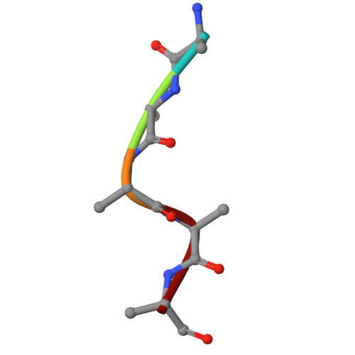
Unidentified MIB1 peptideC [auth D]5Homo sapiensMutation(s): 0 
Gene Names: MIB1
Sequence Annotations
Expand
  • Reference Sequence
Experimental Data & Validation

Experimental Data

  • Method: X-RAY DIFFRACTION
  • Resolution: 2.39 Å
  • R-Value Free: 0.244 
  • R-Value Work: 0.207 
  • R-Value Observed: 0.208 
  • Space Group: C 1 2 1
Unit Cell:
Length ( Å )Angle ( ˚ )
a = 78.997α = 90
b = 137.47β = 108.14
c = 96.248γ = 90
Software Package:
Software NamePurpose
PHENIXrefinement
HKL-2000data reduction
HKL-2000data scaling
PHASERphasing
Cootmodel building

Structure Validation

View Full Validation Report


Entry History & Funding Information

Deposition Data


Funding OrganizationLocationGrant Number
National Institutes of Health/National Cancer Institute (NIH/NCI)United StatesR35 CA220340

Revision History  (Full details and data files)

  • Version 1.0: 2024-09-25
    Type: Initial release