8UXQ | pdb_00008uxq

Structure of Heterochromatin Protein 1 (HP1) alpha in complex with an H2A.Z nucleosome


Experimental Data Snapshot

  • Method: ELECTRON MICROSCOPY
  • Resolution: 6.30 Å
  • Aggregation State: PARTICLE 
  • Reconstruction Method: SINGLE PARTICLE 

Starting Model: in silico
View more details

wwPDB Validation   3D Report Full Report


This is version 1.1 of the entry. See complete history


Literature

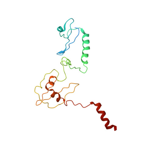
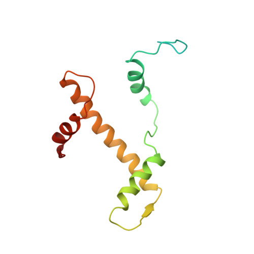
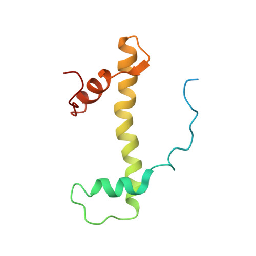
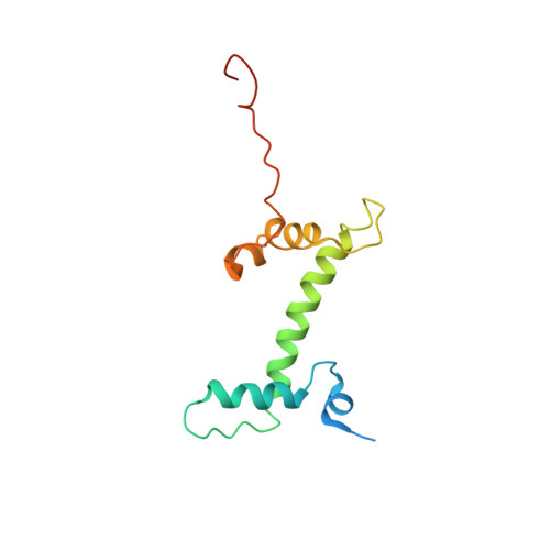
Structural mechanism of HP1⍺-dependent transcriptional repression and chromatin compaction.

Sokolova, V.Miratsky, J.Svetlov, V.Brenowitz, M.Vant, J.Lewis, T.S.Dryden, K.Lee, G.Sarkar, S.Nudler, E.Singharoy, A.Tan, D.

(2024) Structure 32: 2094-2106.e6

  • DOI: https://doi.org/10.1016/j.str.2024.09.013
  • Primary Citation of Related Structures:  
    8UXQ

  • PubMed Abstract: 

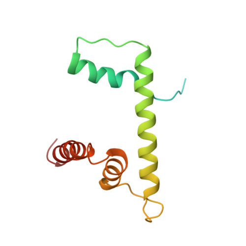
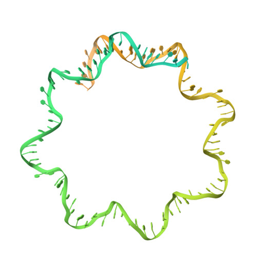
    Heterochromatin protein 1 (HP1) plays a central role in establishing and maintaining constitutive heterochromatin. However, the mechanisms underlying HP1-nucleosome interactions and their contributions to heterochromatin functions remain elusive. Here, we present the cryoelectron microscopy (cryo-EM) structure of an HP1α dimer bound to an H2A.Z-nucleosome, revealing two distinct HP1α-nucleosome interfaces. The primary HP1α binding site is located at the N terminus of histone H3, specifically at the trimethylated lysine 9 (K9me3) region, while a secondary binding site is situated near histone H2B, close to nucleosome superhelical location 4 (SHL4). Our biochemical data further demonstrates that HP1α binding influences the dynamics of DNA on the nucleosome. It promotes DNA unwrapping near the nucleosome entry and exit sites while concurrently restricting DNA accessibility in the vicinity of SHL4. Our study offers a model for HP1α-mediated heterochromatin maintenance and gene silencing. It also sheds light on the H3K9me-independent role of HP1 in responding to DNA damage.


  • Organizational Affiliation
    • Department of Pharmacological Sciences, Stony Brook University, Stony Brook, NY, USA.

Macromolecules

Find similar proteins by:  (by identity cutoff)  |  3D Structure
Entity ID: 1
MoleculeChains Sequence LengthOrganismDetailsImage
Chromobox protein homolog 5A,
B [auth C]
195Homo sapiensMutation(s): 0 
Gene Names: CBX5HP1A
UniProt & NIH Common Fund Data Resources
Find proteins for P45973 (Homo sapiens)
Explore P45973 
Go to UniProtKB:  P45973
PHAROS:  P45973
GTEx:  ENSG00000094916 
Entity Groups  
Sequence Clusters30% Identity50% Identity70% Identity90% Identity95% Identity100% Identity
UniProt GroupP45973
Sequence Annotations
Expand
  • Reference Sequence
Find similar proteins by:  (by identity cutoff)  |  3D Structure
Entity ID: 2
MoleculeChains Sequence LengthOrganismDetailsImage
Histone H3.2C [auth E],
G [auth K]
135Xenopus laevisMutation(s): 1 
UniProt
Find proteins for P84233 (Xenopus laevis)
Explore P84233 
Go to UniProtKB:  P84233
Entity Groups  
Sequence Clusters30% Identity50% Identity70% Identity90% Identity95% Identity100% Identity
UniProt GroupP84233
Sequence Annotations
Expand
  • Reference Sequence
Find similar proteins by:  (by identity cutoff)  |  3D Structure
Entity ID: 3
MoleculeChains Sequence LengthOrganismDetailsImage
Histone H4D [auth F],
H [auth L]
102Xenopus laevisMutation(s): 0 
UniProt
Find proteins for P62799 (Xenopus laevis)
Explore P62799 
Go to UniProtKB:  P62799
Entity Groups  
Sequence Clusters30% Identity50% Identity70% Identity90% Identity95% Identity100% Identity
UniProt GroupP62799
Sequence Annotations
Expand
  • Reference Sequence
Find similar proteins by:  (by identity cutoff)  |  3D Structure
Entity ID: 4
MoleculeChains Sequence LengthOrganismDetailsImage
Histone H2A.ZE [auth G],
I [auth M]
127Mus musculusMutation(s): 0 
Gene Names: H2az1H2afzH2az
UniProt & NIH Common Fund Data Resources
Find proteins for P0C0S6 (Mus musculus)
Explore P0C0S6 
Go to UniProtKB:  P0C0S6
IMPC:  MGI:1888388
Entity Groups  
Sequence Clusters30% Identity50% Identity70% Identity90% Identity95% Identity100% Identity
UniProt GroupP0C0S6
Sequence Annotations
Expand
  • Reference Sequence
Find similar proteins by:  (by identity cutoff)  |  3D Structure
Entity ID: 5
MoleculeChains Sequence LengthOrganismDetailsImage
Histone H2B 1.1F [auth H],
J [auth N]
125Xenopus laevisMutation(s): 0 
UniProt
Find proteins for P02281 (Xenopus laevis)
Explore P02281 
Go to UniProtKB:  P02281
Entity Groups  
Sequence Clusters30% Identity50% Identity70% Identity90% Identity95% Identity100% Identity
UniProt GroupP02281
Sequence Annotations
Expand
  • Reference Sequence
Find similar nucleic acids by:  (by identity cutoff)  |  3D Structure
Entity ID: 6
MoleculeChains LengthOrganismImage
DNA Widom601 (208bp) strand1K [auth I]208synthetic construct
Sequence Annotations
Expand
  • Reference Sequence
Find similar nucleic acids by:  (by identity cutoff)  |  3D Structure
Entity ID: 7
MoleculeChains LengthOrganismImage
DNA Widom601 (208bp) strand2L [auth J]208synthetic construct
Sequence Annotations
Expand
  • Reference Sequence
Experimental Data & Validation

Experimental Data

  • Method: ELECTRON MICROSCOPY
  • Resolution: 6.30 Å
  • Aggregation State: PARTICLE 
  • Reconstruction Method: SINGLE PARTICLE 
EM Software:
TaskSoftware PackageVersion
MODEL REFINEMENTMDFF
RECONSTRUCTIONRELION4.0

Structure Validation

View Full Validation Report


Entry History & Funding Information

Deposition Data


Funding OrganizationLocationGrant Number
National Institutes of Health/National Institute of General Medical Sciences (NIH/NIGMS)United StatesR35GM133611
National Science Foundation (NSF, United States)United States1942049

Revision History  (Full details and data files)

  • Version 1.0: 2024-09-25
    Type: Initial release
  • Version 1.1: 2025-04-09
    Changes: Data collection, Database references, Structure summary