A domain-swapped CaMKII conformation facilitates linker-mediated allosteric regulation.
Nguyen, B.V., Ozden, C., Dong, K., Koc, O.C., Torres-Ocampo, A.P., Dziedzic, N., Flaherty, D., Huang, J., Sankara, S., Abromson, N.L., Tomchick, D.R., Fissore, R.A., Chen, J., Garman, S.C., Stratton, M.M.(2025) Nat Commun 16: 8461-8461
- PubMed: 41006217 Search on PubMedSearch on PubMed Central
- DOI: https://doi.org/10.1038/s41467-025-63249-w
- Primary Citation Related Structures:
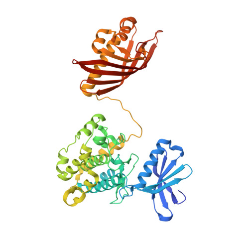
8USO - PubMed Abstract:
Memory formation, fertilization, and cardiac function rely on precise Ca 2+ signaling and subsequent Ca 2+ /calmodulin-dependent protein kinase II (CaMKII) activation. Ca 2+ sensitivity of the four CaMKII paralogs in mammals is linked to the length of the variable linker region that undergoes extensive alternative splicing. In this study, we determine that the position of charged residues within the linker modulates the Ca 2+ /CaM sensitivity. We present an X-ray crystal structure of the full-length CaMKIIδ holoenzyme consisting of domain-swapped dimers within a dodecameric complex, revealing potential contacts for cooperativity and allostery. Based on molecular dynamics (MD) simulations, small-angle X-ray scattering (SAXS) measurements, and live-cell imaging, we propose a model where the domain-swapped conformation positions the charges of the linker region to drive an interaction with the regulatory segment that modulates the degree of autoinhibition. Our findings provide a framework for understanding allosteric regulation of CaMKII by the linker region in Ca 2+ -sensitive cells.
- Molecular and Cellular Biology Graduate Program, University of Massachusetts, Amherst, MA, USA.
Organizational Affiliation: