8URN

Crystal structure of EscI(51-87)-linker-EtgA(18-152) fusion protein

  • Classification: HYDROLASE
  • Organism(s): Escherichia coli
  • Expression System: Escherichia coli
  • Mutation(s): No 

  • Deposited: 2023-10-26 Released: 2024-02-14 
  • Deposition Author(s): van den Akker, F.
  • Funding Organization(s): National Institutes of Health/National Institute Of Allergy and Infectious Diseases (NIH/NIAID)

Experimental Data Snapshot

  • Method: X-RAY DIFFRACTION
  • Resolution: 2.01 Å
  • R-Value Free: 0.245 
  • R-Value Work: 0.189 
  • R-Value Observed: 0.191 

wwPDB Validation   3D Report Full Report


This is version 1.1 of the entry. See complete history


Literature

Structural insights into peptidoglycan glycosidase EtgA binding to the inner rod protein EscI of the type III secretion system via a designed EscI-EtgA fusion protein.

Boorman, J.Zeng, X.Lin, J.van den Akker, F.

(2024) Protein Sci 33: e4930-e4930

  • DOI: https://doi.org/10.1002/pro.4930
  • Primary Citation of Related Structures:  
    8URN

  • PubMed Abstract: 

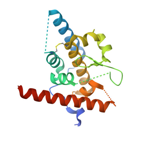
    Bacteria express lytic enzymes such as glycosidases, which have potentially self-destructive peptidoglycan (PG)-degrading activity and, therefore, require careful regulation in bacteria. The PG glycosidase EtgA is regulated by localization to the assembling type III secretion system (T3SS), generating a hole in the PG layer for the T3SS to reach the outer membrane. The EtgA localization was found to be mediated via EtgA interacting with the T3SS inner rod protein EscI. To gain structural insights into the EtgA recognition of EscI, we determined the 2.01 Å resolution structure of an EscI (51-87)-linker-EtgA fusion protein designed based on AlphaFold2 predictions. The structure revealed EscI residues 72-87 forming an α-helix interacting with the backside of EtgA, distant from the active site. EscI residues 56-71 also were found to interact with EtgA, with these residues stretching across the EtgA surface. The ability of the EscI to interact with EtgA was also probed using an EscI peptide. The EscI peptide comprising residues 66-87, slightly larger than the observed EscI α-helix, was shown to bind to EtgA using microscale thermophoresis and thermal shift differential scanning fluorimetry. The EscI peptide also had a two-fold activity-enhancing effect on EtgA, whereas the EscI-EtgA fusion protein enhanced activity over four-fold compared to EtgA. Our studies suggest that EtgA regulation by EscI could be trifold involving protein localization, protein activation, and protein stabilization components. Analysis of the sequence conservation of the EscI EtgA interface residues suggested a possible conservation of such regulation for related proteins from different bacteria.


  • Organizational Affiliation

    Department of Biochemistry, Case Western Reserve University, Cleveland, Ohio, USA.


Macromolecules
Find similar proteins by:  (by identity cutoff)  |  3D Structure
Entity ID: 1
MoleculeChains Sequence LengthOrganismDetailsImage
EscI inner rod protein type III secretion system,EtgA protein189Escherichia coliMutation(s): 0 
Gene Names: escIC9E67_00595etgA
UniProt
Find proteins for O85634 (Escherichia coli)
Explore O85634 
Go to UniProtKB:  O85634
Find proteins for C7BUG6 (Escherichia coli)
Explore C7BUG6 
Go to UniProtKB:  C7BUG6
Entity Groups  
Sequence Clusters30% Identity50% Identity70% Identity90% Identity95% Identity100% Identity
UniProt GroupsC7BUG6O85634
Sequence Annotations
Expand
  • Reference Sequence
Small Molecules
Ligands 1 Unique
IDChains Name / Formula / InChI Key2D Diagram3D Interactions
SO4
Query on SO4

Download Ideal Coordinates CCD File 
B [auth A]SULFATE ION
O4 S
QAOWNCQODCNURD-UHFFFAOYSA-L
Experimental Data & Validation

Experimental Data

  • Method: X-RAY DIFFRACTION
  • Resolution: 2.01 Å
  • R-Value Free: 0.245 
  • R-Value Work: 0.189 
  • R-Value Observed: 0.191 
  • Space Group: C 1 2 1
Unit Cell:
Length ( Å )Angle ( ˚ )
a = 107.629α = 90
b = 29.885β = 118.19
c = 74.564γ = 90
Software Package:
Software NamePurpose
REFMACrefinement
XDSdata scaling
XDSdata reduction
PHASERphasing

Structure Validation

View Full Validation Report


Entry History & Funding Information

Deposition Data


Funding OrganizationLocationGrant Number
National Institutes of Health/National Institute Of Allergy and Infectious Diseases (NIH/NIAID)United States1R21AI148875

Revision History  (Full details and data files)

  • Version 1.0: 2024-02-14
    Type: Initial release
  • Version 1.1: 2024-03-20
    Changes: Database references