8URA | pdb_00008ura

Crystal structure of Corynebacterium ulcerans endo-beta-N-acetylglucosaminidase catalytically inactive CU43 D187A-E189A at 2.6 A resolution (space group P21)


Experimental Data Snapshot

  • Method: X-RAY DIFFRACTION
  • Resolution: 2.59 Å
  • R-Value Free: 
    0.249 (Depositor), 0.249 (DCC) 
  • R-Value Work: 
    0.191 (Depositor), 0.192 (DCC) 
  • R-Value Observed: 
    0.197 (Depositor) 

Starting Model: in silico
View more details

wwPDB Validation 3D Report Full Report

Validation slider image for 8URA

This is version 1.2 of the entry. See complete history

Literature

Potent efficacy of an IgG-specific endoglycosidase against IgG-mediated pathologies.

Sastre, D.E.Bournazos, S.Du, J.Boder, E.J.Edgar, J.E.Azzam, T.Sultana, N.Huliciak, M.Flowers, M.Yoza, L.Xu, T.Chernova, T.A.Ravetch, J.V.Sundberg, E.J.

(2024) Cell 187: 6994

  • DOI: https://doi.org/10.1016/j.cell.2024.09.038
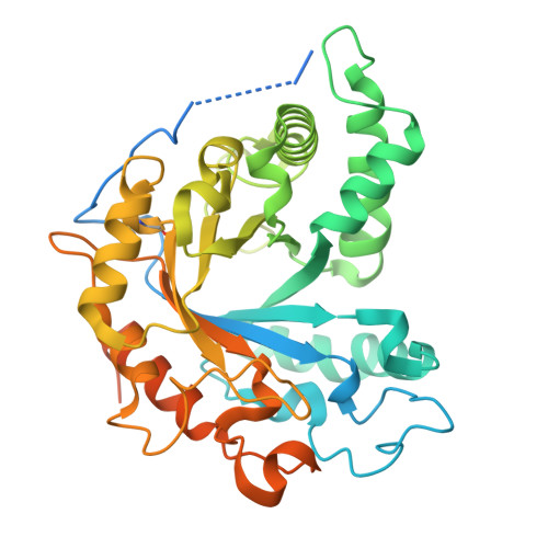
  • Primary Citation Related Structures: 
    8UEN, 8URA

  • PubMed Abstract: 

    Endo-β-N-acetylglucosaminidases (ENGases) that specifically hydrolyze the Asn297-linked glycan on immunoglobulin G (IgG) antibodies, the major molecular determinant of fragment crystallizable (Fc) γ receptor (FcγR) binding, are exceedingly rare. All previously characterized IgG-specific ENGases are multi-domain proteins secreted as an immune evasion strategy by Streptococcus pyogenes strains. Here, using in silico analysis and mass spectrometry techniques, we identified a family of single-domain ENGases secreted by pathogenic corynebacterial species that exhibit strict specificity for IgG antibodies. By X-ray crystallographic and surface plasmon resonance analyses, we found that the most catalytically efficient IgG-specific ENGase family member recognizes both protein and glycan components of IgG. Employing in vivo models, we demonstrated the remarkable efficacy of this IgG-specific ENGase in mitigating numerous pathologies that rely on FcγR-mediated effector functions, including T and B lymphocyte depletion, autoimmune hemolytic anemia, and antibody-dependent enhancement of dengue disease, revealing its potential for treating and/or preventing a wide range of IgG-mediated diseases in humans.


  • Organizational Affiliation
    • Department of Biochemistry, Emory University School of Medicine, Atlanta, GA 30322, USA. Electronic address: dsastre@emory.edu.

Macromolecule Content 

  • Total Structure Weight: 92.21 kDa 
  • Atom Count: 5,145 
  • Modeled Residue Count: 647 
  • Deposited Residue Count: 830 
  • Unique protein chains: 1

Macromolecules

Find similar proteins by:|  3D Structure
Entity ID: 1
MoleculeChains  Sequence LengthOrganismDetailsImage
Corynebacterial protease CP40
A, B
415Corynebacterium ulceransMutation(s): 2 
Gene Names: cppCULC0211_20780

Experimental Data & Validation

Experimental Data

  • Method: X-RAY DIFFRACTION
  • Resolution: 2.59 Å
  • R-Value Free:  0.249 (Depositor), 0.249 (DCC) 
  • R-Value Work:  0.191 (Depositor), 0.192 (DCC) 
  • R-Value Observed: 0.197 (Depositor) 
Space Group: P 1 21 1
Unit Cell:
Length ( Å )Angle ( ˚ )
a = 58.514α = 90
b = 70.897β = 92.53
c = 73.734γ = 90
Software Package:
Software NamePurpose
PHENIXrefinement
HKL-2000data reduction
HKL-2000data scaling
PHASERphasing

Structure Validation

View Full Validation Report


Entry History 

& Funding Information

Deposition Data


Funding OrganizationLocationGrant Number
National Institutes of Health/National Institute Of Allergy and Infectious Diseases (NIH/NIAID)United States--

Revision History  (Full details and data files)

  • Version 1.0: 2024-10-23
    Type: Initial release
  • Version 1.1: 2024-11-06
    Changes: Database references
  • Version 1.2: 2024-12-11
    Changes: Database references