8UPI | pdb_00008upi

Structure of a periplasmic peptide binding protein from Mesorhizobium sp. AP09 bound to aminoserine


Experimental Data Snapshot

  • Method: X-RAY DIFFRACTION
  • Resolution: 1.55 Å
  • R-Value Free: 
    0.229 (Depositor), 0.228 (DCC) 
  • R-Value Work: 
    0.179 (Depositor), 0.179 (DCC) 
  • R-Value Observed: 
    0.181 (Depositor) 

Starting Model: in silico
View more details

wwPDB Validation 3D Report Full Report

Validation slider image for 8UPI

Ligand Structure Quality Assessment 


This is version 1.3 of the entry. See complete history

Literature

Identification and Characterization of a Bacterial Periplasmic Solute Binding Protein That Binds l-Amino Acid Amides.

Smith, O.B.Frkic, R.L.Rahman, M.G.Jackson, C.J.Kaczmarski, J.A.

(2024) Biochemistry 63: 1322-1334

  • DOI: https://doi.org/10.1021/acs.biochem.4c00096
  • Primary Citation Related Structures: 
    8UPI

  • PubMed Abstract: 

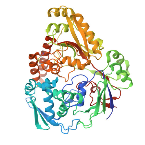
    Periplasmic solute-binding proteins (SBPs) are key ligand recognition components of bacterial ATP-binding cassette (ABC) transporters that allow bacteria to import nutrients and metabolic precursors from the environment. Periplasmic SBPs comprise a large and diverse family of proteins, of which only a small number have been empirically characterized. In this work, we identify a set of 610 unique uncharacterized proteins within the SBP_bac_5 family that are found in conserved operons comprising genes encoding (i) ABC transport systems and (ii) putative amidases from the FmdA_AmdA family. From these uncharacterized SBP_bac_5 proteins, we characterize a representative periplasmic SBP from Mesorhizobium sp. A09 ( Me Ami_SBP) and show that Me Ami_SBP binds l-amino acid amides but not the corresponding l-amino acids. An X-ray crystal structure of Me Ami_SBP bound to l-serinamide highlights the residues that impart distinct specificity for l-amino acid amides and reveals a structural Ca 2+ binding site within one of the lobes of the protein. We show that the residues involved in ligand and Ca 2+ binding are conserved among the 610 SBPs from experimentally uncharacterized FmdA_AmdA amidase-associated ABC transporter systems, suggesting these homologous systems are also likely to be involved in the sensing, uptake, and metabolism of l-amino acid amides across many Gram-negative nitrogen-fixing soil bacteria. We propose that Me Ami_SBP is involved in the uptake of such solutes to supplement pathways such as the citric acid cycle and the glutamine synthetase-glutamate synthase pathway. This work expands our currently limited understanding of microbial interactions with l-amino acid amides and bacterial nitrogen utilization.


  • Organizational Affiliation
    • Research School of Chemistry, Australian National University, Canberra, Australian Capital Territory 2601, Australia.

Macromolecule Content 

  • Total Structure Weight: 57.56 kDa 
  • Atom Count: 4,948 
  • Modeled Residue Count: 510 
  • Deposited Residue Count: 523 
  • Unique protein chains: 1

Macromolecules

Find similar proteins by:|  3D Structure
Entity ID: 1
MoleculeChains  Sequence LengthOrganismDetailsImage
Peptide ABC transporter substrate-binding protein523Mesorhizobium sp. AP09Mutation(s): 0 
Gene Names: MesoLj113b_06930
UniProt
Find proteins for A0ACD6B9E4 (Mesorhizobium sp. 113-3-3)
Explore A0ACD6B9E4 
Go to UniProtKB:  A0ACD6B9E4
Entity Groups
Sequence Clusters30% Identity50% Identity70% Identity90% Identity95% Identity100% Identity
UniProt GroupA0ACD6B9E4
Sequence Annotations
Expand
Reference Sequence

Experimental Data & Validation

Experimental Data

  • Method: X-RAY DIFFRACTION
  • Resolution: 1.55 Å
  • R-Value Free:  0.229 (Depositor), 0.228 (DCC) 
  • R-Value Work:  0.179 (Depositor), 0.179 (DCC) 
  • R-Value Observed: 0.181 (Depositor) 
Space Group: P 21 21 21
Unit Cell:
Length ( Å )Angle ( ˚ )
a = 56.247α = 90
b = 58.92β = 90
c = 139.355γ = 90
Software Package:
Software NamePurpose
PHENIXrefinement
Aimlessdata scaling
CrysalisProdata reduction
PHASERphasing

Structure Validation

View Full Validation Report


Ligand Structure Quality Assessment 


Entry History 

& Funding Information

Deposition Data


Funding OrganizationLocationGrant Number
Australian Research Council (ARC)AustraliaCE200100029

Revision History  (Full details and data files)

  • Version 1.0: 2023-11-08
    Type: Initial release
  • Version 1.1: 2024-05-22
    Changes: Database references
  • Version 1.2: 2024-06-05
    Changes: Database references
  • Version 1.3: 2024-10-23
    Changes: Structure summary